. 2017 Feb 28;8(9):14462-14478.
doi: 10.18632/oncotarget.14895.

The ribosomal protein gene RPL5 is a haploinsufficient tumor suppressor in multiple cancer types

Affiliations

The ribosomal protein gene RPL5 is a haploinsufficient tumor suppressor in multiple cancer types

Laura Fancello et al. Oncotarget. .

Abstract

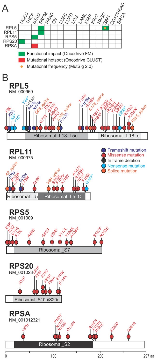
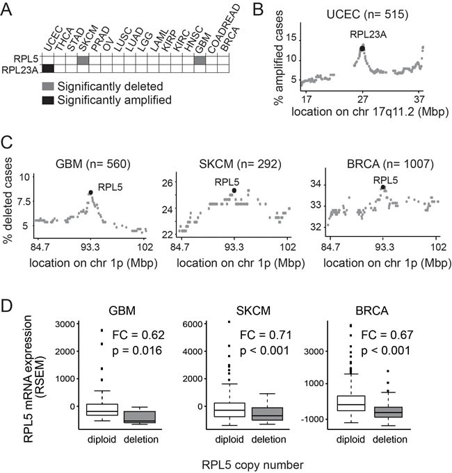
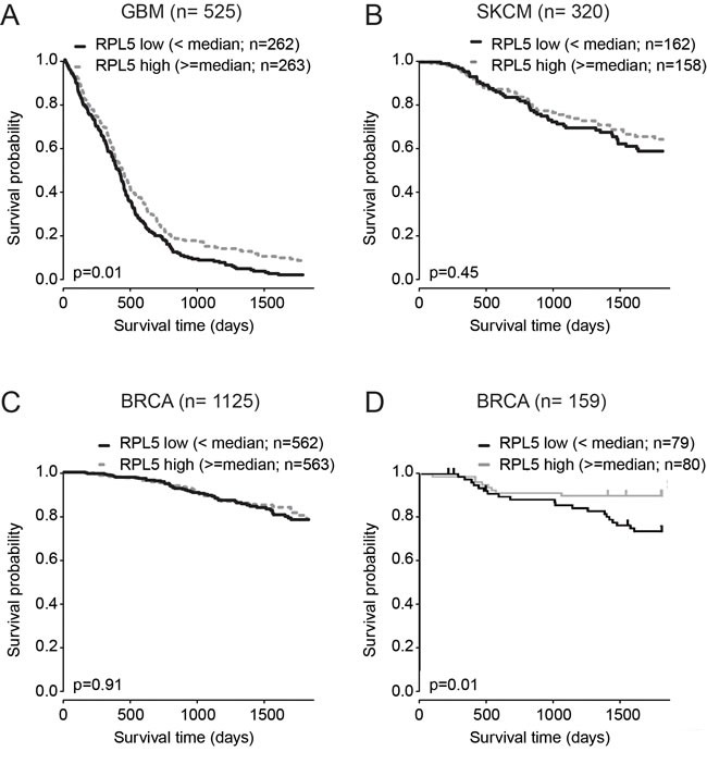
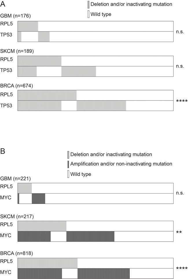
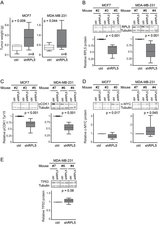
For many years, defects in the ribosome have been associated to cancer. Recently, somatic mutations and deletions affecting ribosomal protein genes were identified in a few leukemias and solid tumor types. However, systematic analysis of all 81 known ribosomal protein genes across cancer types is lacking. We screened mutation and copy number data of respectively 4926 and 7322 samples from 16 cancer types and identified six altered genes (RPL5, RPL11, RPL23A, RPS5, RPS20 and RPSA). RPL5 was located at a significant peak of heterozygous deletion or mutated in 11% of glioblastoma, 28% of melanoma and 34% of breast cancer samples. Moreover, patients with low RPL5 expression displayed worse overall survival in glioblastoma and in one breast cancer cohort. RPL5 knockdown in breast cancer cell lines enhanced G2/M cell cycle progression and accelerated tumor progression in a xenograft mouse model. Interestingly, our data suggest that the tumor suppressor role of RPL5 is not only mediated by its known function as TP53 or c-MYC regulator. In conclusion, RPL5 heterozygous inactivation occurs at high incidence (11-34%) in multiple tumor types, currently representing the most common somatic ribosomal protein defect in cancer, and we demonstrate a tumor suppressor role for RPL5 in breast cancer.

Keywords: TCGA; breast cancer; haploinsufficient tumor suppressor; ribosomal protein.


Conflict of interest statement

CONFLICTs OF INTEREST

There are no conflicts of interest.

Figures

Figure 1
Figure 1. Identification of 5 ribosomal protein genes that are significantly mutated in cancer
(A) Significantly mutated genes identified due to mutational frequency (MutSig 2.0), mutational clustering (OncodriveCLUST) or accumulation of high functional impact mutations (OncodriveFM). (B) Mapping of mutations affecting the 5 candidate cancer drivers on linear protein diagrams. Non-silent somatic mutations from all 16 cancer types are shown. Protein domains are indicated for each protein. Ribosomal_L18_L5e (pfam00861), Ribosomal_L18_c (pfam14204): RPL5 protein domains. Ribosomal_L5 (pfam00281), Ribosomal_L5_C (pfam00673): RPL11 protein domains. Ribosomal_S7 (pfam00177): RPS5 protein domain. Ribosomal_S10p/S20e (pfam00338): RPS20 protein domain. Ribosomal_S2 (pfam00318): RPSA protein domain.
Figure 2
Figure 2. RPL5 and RPL23A show significant copy number changes in the TCGA database
(A) Heatmap showing the significant copy number changes retained (B) RPL23A amplification peak in UCEC. Each dot on the figure represents a different gene on chr 17q11.2, the genomic locus where the RPL23A gene is located. X-axis: genomic coordinates on chromosome 17; Y-axis: percent of cases with amplification of each particular gene. (C) RPL5 deletion peak in GBM, SKCM and BRCA. Each dot on the figure represents a different gene on chr 1p, the genomic locus where the RPL5 gene is located. X-axis: genomic coordinates on chromosome 1; Y-axis: percent of cases with deletion of each particular gene. (D) Boxplots showing RPL5 mRNA expression levels (RSEM) in GBM, SKCM and BRCA cases with diploid or heterozygously deleted copy number status for RPL5. P: p-value according to the Wilcoxon's test. FC: fold change (RPL5 heterozygously deleted over RPL5 diploid).
Figure 3
Figure 3. RPL5 is a clinically relevant candidate tumor suppressor in GBM
Kaplan–Meier analysis of the effect of RPL5 expression on overall survival. Cases were divided in RPL5 low or high expressers according to whether expression was below or above median and survival was compared using the log-rank test. (A) GBM TCGA dataset; (B) SKCM TCGA dataset; (C) BRCA TCGA dataset; (D) Non-TCGA BRCA dataset available on the R2 platform (GEO accession: GSE1456).
Figure 4
Figure 4. Genetic interaction between RPL5 alterations and TP53 or c-MYC alterations
(A) Co-occurrence of RPL5 and TP53 alterations in GBM, SKCM and BRCA. (B) Co-occurrence of RPL5 and c-MYC alterations in GBM, SKCM and BRCA.
Figure 5
Figure 5. RPL5 knockdown enhances proliferation of MCF7 breast cancer cells
(A) Immunoblot analysis of RPL5 expression levels on cell lysates of doxycycline treated MCF7 and MDA-MB-231 cell lines containing an empty lentiviral vector (control) or a vector containing an shRNA targeting RPL5 (shRPL5). Quantification of the blots is shown on the right. (B) In vitro proliferation of MCF7 and MDA-MB-231 cell lines as determined by real-time monitoring of cell confluency. (C) Immunoblot analysis of c-MYC, TP53 and MDM2 expression levels on cell lysates of doxycycline treated MCF7 and MDA-MB-231 cell lines containing an empty lentiviral vector (control) or a vector containing an shRNA targeting RPL5 (shRPL5). All immunoblots were performed 72 hrs after start of the doxycycline treatment.
Figure 6
Figure 6. Knockdown of RPL5 accelerates breast cancer formation in mice
(A) Tumor weights of doxycycline treated mice that are injected with MCF7 and MDA-MB-231 cell lines containing the empty lentiviral vector (control) in the left flanks and the vector containing an shRNA targeting RPL5 (shRPL5) in the rights flanks. (B-E) Immunoblot analysis and the corresponding quantification of MCF7 (left) and MDA-MB-231 (right) tumors, comparing expression of the control and shRPL5 condition for RPL5 (B), phospho-CDK1 (tyr15) (C), c-MYC (D), and TP53 protein (E).

References

    1. De Keersmaecker K, Atak ZK, Li N, Vicente C, Patchett S, Girardi T, Gianfelici V, Geerdens E, Clappier E, Porcu M, Lahortiga I, Lucà R, Yan J, et al. Exome sequencing identifies mutation in CNOT3 and ribosomal genes RPL5 and RPL10 in T-cell acute lymphoblastic leukemia. Nat Genet. 2013;45:186–90. doi: 10.1038/ng.2508. - DOI - PMC - PubMed
    1. Rao S, Lee SY, Gutierrez A, Perrigoue J, Thapa RJ, Tu Z, Jeffers JR, Rhodes M, Anderson S, Oravecz T, Hunger SP, Timakhov RA, Zhang R, et al. Inactivation of ribosomal protein L22 promotes transformation by induction of the stemness factor, Lin28B. Blood. 2012;120:3764–73. doi: 10.1182/blood-2012-03-415349. - DOI - PMC - PubMed
    1. Tzoneva G, Perez-Garcia A, Carpenter Z, Khiabanian H, Tosello V, Allegretta M, Paietta E, Racevskis J, Rowe JM, Tallman MS, Paganin M, Basso G, Hof J, et al. Activating mutations in the NT5C2 nucleotidase gene drive chemotherapy resistance in relapsed ALL. Nat Med. 2013;19:368–71. doi: 10.1038/nm.3078. - DOI - PMC - PubMed
    1. Landau D a, Tausch E, Taylor-Weiner AN, Stewart C, Reiter JG, Bahlo J, Kluth S, Bozic I, Lawrence M, Böttcher S, Carter SL, Cibulskis K, Mertens D, et al. Mutations driving CLL and their evolution in progression and relapse. Nature. 2015. - DOI - PMC - PubMed
    1. Ljungström V, Cortese D, Young E, Pandzic T, Mansouri L, Plevova K, Ntoufa S, Baliakas P, Clifford R, Sutton LA, Blakemore SJ, Stavroyianni N, Agathangelidis A, et al. Whole-exome sequencing in relapsing chronic lymphocytic leukemia: Clinical impact of recurrent RPS15 mutations. Blood. 2016;127:1007–16. doi: 10.1182/blood-2015-10-674572. - DOI - PMC - PubMed