Review
. 2017 Feb 2;19(1):18.
doi: 10.1186/s13075-017-1229-9.

Synovitis in osteoarthritis: current understanding with therapeutic implications

Affiliations
Review

Synovitis in osteoarthritis: current understanding with therapeutic implications

Alexander Mathiessen et al. Arthritis Res Ther. .

Abstract

Modern concepts of osteoarthritis (OA) have been forever changed by modern imaging phenotypes demonstrating complex and multi-tissue pathologies involving cartilage, subchondral bone and (increasingly recognized) inflammation of the synovium. The synovium may show significant changes, even before visible cartilage degeneration has occurred, with infiltration of mononuclear cells, thickening of the synovial lining layer and production of inflammatory cytokines. The combination of sensitive imaging modalities and tissue examination has confirmed a high prevalence of synovial inflammation in all stages of OA, with a number of studies demonstrating that synovitis is related to pain, poor function and may even be an independent driver of radiographic OA onset and structural progression. Treating key aspects of synovial inflammation therefore holds great promise for analgesia and also for structure modification. This article will review current knowledge on the prevalence of synovitis in OA and its role in symptoms and structural progression, and explore lessons learnt from targeting synovitis therapeutically.

Keywords: Epidemiology; Imaging; Osteoarthritis; Pathophysiology; Synovitis; Treatment.


Figures
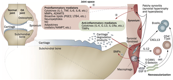
Fig. 1
Fig. 1
Involvement of the synovium in OA pathophysiology. Products of cartilage breakdown released into the synovial fluid are phagocytosed by synovial cells, amplifying synovial inflammation. In turn, activated synovial cells in the inflamed synovium produce catabolic and pro-inflammatory mediators that lead to excess production of the proteolytic enzymes responsible for cartilage breakdown, creating a positive feedback loop. The inflammatory response is amplified by activated synovial T cells, B cells and infiltrating macrophages. To counteract this inflammatory response, the synovium and cartilage may produce anti-inflammatory cytokines. In addition to these effects on cartilage inflammation and breakdown, the inflamed synovium contributes to the formation of osteophytes via BMPs. ADAMTS a disintegrin and metalloproteinase with thrombospondin motifs, BMP bone morphogenetic protein, CXCL13 CXC-chemokine ligand 13, EGF endothelial growth factor, IL interleukin, IL-1Ra IL-1 receptor antagonist, LTB4, leukotriene B4, MMP matrix metalloproteinase, NAMPT nicotinamide phosphoribosyl transferase (visfatin), NO nitric oxide, OA osteoarthritis, PGE2 prostaglandin E2, TiMP tissue inhibitor of metalloproteinase, TNF tumour necrosis factor, VCAM-1, vascular cell adhesion molecule 1, VEGF vascular endothelial growth factor (Reprinted from [5] with permission from Macmillan Publishers Ltd)
Fig. 2
Fig. 2
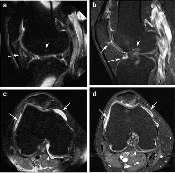
a Sagittal fat-suppressed proton density-weighted image (non-CE-MRI) shows hyperintensity in the intercondylar (arrow) region of Hoffa’s fat pad. This signal alteration is used as a surrogate marker for synovitis on non-CE-MRI. There is a discrete subchondral bone marrow alteration in the femur (arrowhead) corresponding to the site of the anterior cruciate ligament (ACL) insertion. b In contrast, sagittal fat-suppressed T1-weighted contrast-enhanced MRI (CE-MRI) shows no intercondylar synovitis, but reveals infrapatellar synovitis and synovitis adjacent to the tibial ACL insertion (arrows) not seen on non-CE-MRI, as well as bone marrow edema (arrowhead). c Axial non-CE-MRI shows a fluid-equivalent signal within the joint cavity suggestive of joint effusion in the peripatellar recesses (arrows) and posteriorly (arrowhead). d Axial CE-MRI shows marked synovitis anteriorly (arrows) and true effusion only depicted posteriorly as hypointensity adjacent to the synovial lining (arrowhead). Reprinted from [62] with permission from Elsevier

Similar articles

Cited by

References

    1. Division UNDoEaSAP. World Population Prospects: The 2015 Revision, Key Findings and Advance Tables. 2016. https://esa.un.org/unpd/wpp/.
    1. Thomas E, Peat G, Croft P. Defining and mapping the person with osteoarthritis for population studies and public health. Rheumatology (Oxford) 2014;53(2):338–345. doi: 10.1093/rheumatology/ket346. - DOI - PMC - PubMed
    1. Goldenberg DL, Egan MS, Cohen AS. Inflammatory synovitis in degenerative joint disease. J Rheumatol. 1982;9(2):204–209. - PubMed
    1. Berenbaum F. Osteoarthritis as an inflammatory disease (osteoarthritis is not osteoarthrosis!) Osteoarthritis Cartilage. 2013;21(1):16–21. doi: 10.1016/j.joca.2012.11.012. - DOI - PubMed
    1. Sellam J, Berenbaum F. The role of synovitis in pathophysiology and clinical symptoms of osteoarthritis. Nat Rev Rheumatol. 2010;6(11):625–635. doi: 10.1038/nrrheum.2010.159. - DOI - PubMed

MeSH terms