

TBK1: a new player in ALS linking autophagy and neuroinflammation
- PMID: 28148298
- PMCID: PMC5288885
- DOI: 10.1186/s13041-017-0287-x
TBK1: a new player in ALS linking autophagy and neuroinflammation
Abstract
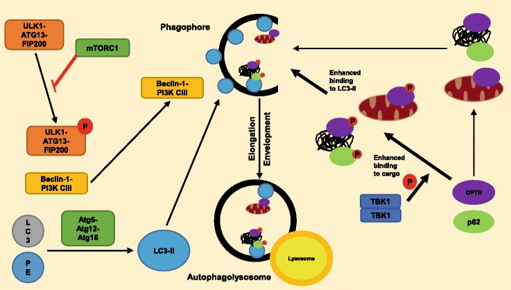
Amyotrophic lateral sclerosis (ALS) is an adult-onset neurodegenerative disorder affecting motor neurons, resulting in progressive muscle weakness and death by respiratory failure. Protein and RNA aggregates are a hallmark of ALS pathology and are thought to contribute to ALS by impairing axonal transport. Mutations in several genes known to contribute to ALS result in deposition of their protein products as aggregates; these include TARDBP, C9ORF72, and SOD1. In motor neurons, this can disrupt transport of mitochondria to areas of metabolic need, resulting in damage to cells and can elicit a neuroinflammatory response leading to further neuronal damage. Recently, eight independent human genetics studies have uncovered a link between TANK-binding kinase 1 (TBK1) mutations and ALS. TBK1 belongs to the IKK-kinase family of kinases that are involved in innate immunity signaling pathways; specifically, TBK1 is an inducer of type-1 interferons. TBK1 also has a major role in autophagy and mitophagy, chiefly the phosphorylation of autophagy adaptors. Several other ALS genes are also involved in autophagy, including p62 and OPTN. TBK1 is required for efficient cargo recruitment in autophagy; mutations in TBK1 may result in impaired autophagy and contribute to the accumulation of protein aggregates and ALS pathology. In this review, we focus on the role of TBK1 in autophagy and the contributions of this process to the pathophysiology of ALS.
Keywords: ALS; Amyotrophic lateral sclerosis; Autophagy; FTD; Frontotemporal dementia; Mitophagy; Motor neuron disease; Neuroinflammation; Signaling; TBK1.
Figures
Similar articles
-
ALS- and FTD-associated missense mutations in TBK1 differentially disrupt mitophagy.Proc Natl Acad Sci U S A. 2021 Jun 15;118(24):e2025053118. doi: 10.1073/pnas.2025053118. Proc Natl Acad Sci U S A. 2021. PMID: 34099552 Free PMC article.
-
Deletion of Tbk1 disrupts autophagy and reproduces behavioral and locomotor symptoms of FTD-ALS in mice.Aging (Albany NY). 2019 Apr 30;11(8):2457-2476. doi: 10.18632/aging.101936. Aging (Albany NY). 2019. PMID: 31039129 Free PMC article.
-
Retinoic acid worsens ATG10-dependent autophagy impairment in TBK1-mutant hiPSC-derived motoneurons through SQSTM1/p62 accumulation.Autophagy. 2019 Oct;15(10):1719-1737. doi: 10.1080/15548627.2019.1589257. Epub 2019 Apr 2. Autophagy. 2019. PMID: 30939964 Free PMC article.
-
Functional and structural consequences of TBK1 missense variants in frontotemporal lobar degeneration and amyotrophic lateral sclerosis.Neurobiol Dis. 2022 Nov;174:105859. doi: 10.1016/j.nbd.2022.105859. Epub 2022 Sep 13. Neurobiol Dis. 2022. PMID: 36113750 Review.
-
Association between TBK1 mutations and risk of amyotrophic lateral sclerosis/frontotemporal dementia spectrum: a meta-analysis.Neurol Sci. 2018 May;39(5):811-820. doi: 10.1007/s10072-018-3246-0. Epub 2018 Jan 18. Neurol Sci. 2018. PMID: 29349657 Review.
Cited by
-
Molecular and Cellular Mechanisms Affected in ALS.J Pers Med. 2020 Aug 25;10(3):101. doi: 10.3390/jpm10030101. J Pers Med. 2020. PMID: 32854276 Free PMC article. Review.
-
Epigenetics in the formation of pathological aggregates in amyotrophic lateral sclerosis.Front Mol Neurosci. 2024 Sep 3;17:1417961. doi: 10.3389/fnmol.2024.1417961. eCollection 2024. Front Mol Neurosci. 2024. PMID: 39290830 Free PMC article. Review.
-
Mitophagy and Neurodegeneration: Between the Knowns and the Unknowns.Front Cell Dev Biol. 2022 Mar 22;10:837337. doi: 10.3389/fcell.2022.837337. eCollection 2022. Front Cell Dev Biol. 2022. PMID: 35392168 Free PMC article. Review.
-
Autophagy and disease: unanswered questions.Cell Death Differ. 2020 Mar;27(3):858-871. doi: 10.1038/s41418-019-0480-9. Epub 2020 Jan 3. Cell Death Differ. 2020. PMID: 31900427 Free PMC article. Review.
-
Immune Signaling Kinases in Amyotrophic Lateral Sclerosis (ALS) and Frontotemporal Dementia (FTD).Int J Mol Sci. 2021 Dec 10;22(24):13280. doi: 10.3390/ijms222413280. Int J Mol Sci. 2021. PMID: 34948077 Free PMC article. Review.
References
Publication types
MeSH terms
Substances
LinkOut - more resources
Full Text Sources
Other Literature Sources
Medical
Miscellaneous
