. 2017 Feb;6(2):73-81.
doi: 10.1302/2046-3758.62.BJR-2016-0199.R1.

Characterisation of osteophytes as an autologous bone graft source: An experimental study in vivo and in vitro

Affiliations

Characterisation of osteophytes as an autologous bone graft source: An experimental study in vivo and in vitro

K Ishihara et al. Bone Joint Res. 2017 Feb.

Abstract

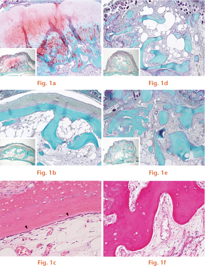
Objectives: Osteophytes are products of active endochondral and intramembranous ossification, and therefore could theoretically provide significant efficacy as bone grafts. In this study, we compared the bone mineralisation effectiveness of osteophytes and cancellous bone, including their effects on secretion of growth factors and anabolic effects on osteoblasts.

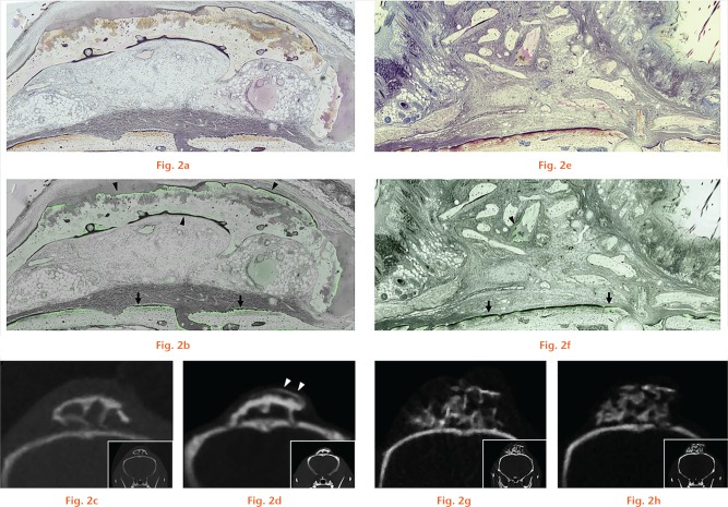
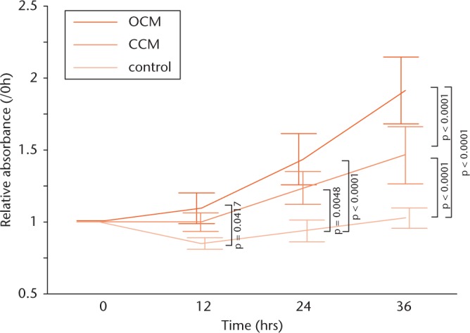
Methods: Osteophytes and cancellous bone obtained from human patients were transplanted onto the calvaria of severe combined immunodeficient mice, with Calcein administered intra-peritoneally for fluorescent labelling of bone mineralisation. Conditioned media were prepared using osteophytes and cancellous bone, and growth factor concentration and effects of each graft on proliferation, differentiation and migration of osteoblastic cells were assessed using enzyme-linked immunosorbent assays, MTS ((3-(4,5-dimethylthiazol-2-yl)-5-(3-carboxymethoxyphenyl)-2-(4-sulfophenyl)-2H-tetrazolium)) assays, quantitative real-time polymerase chain reaction, and migration assays.

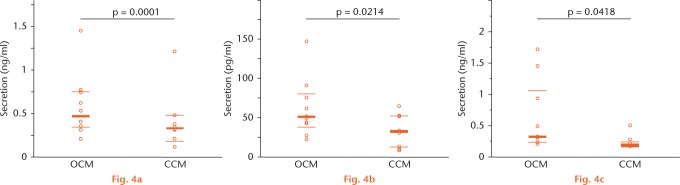
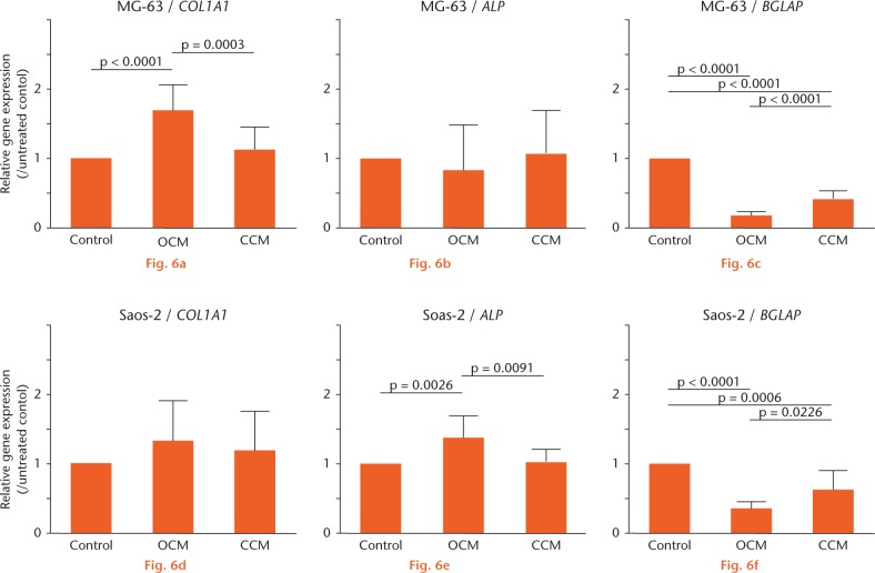
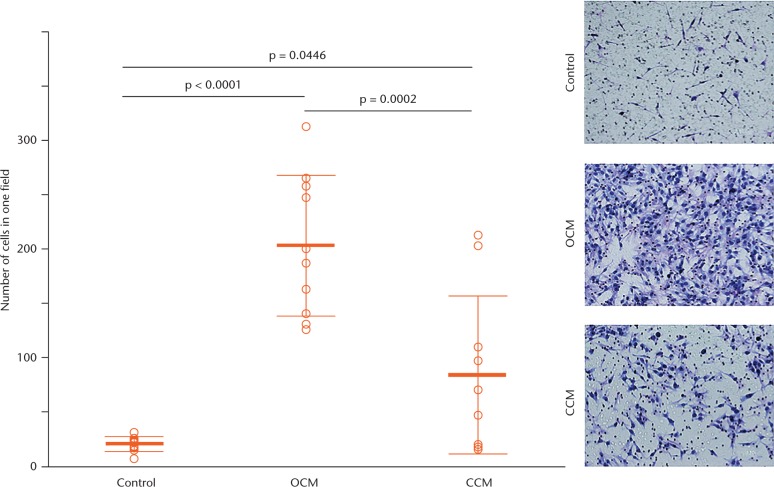
Results: After six weeks, the area of mineralisation was significantly higher for the transplanted osteophytes than for the cancellous bone (43803 μm2, sd 14660 versus 9421 μm2, sd 5032, p = 0.0184, one-way analysis of variance). Compared with cancellous bone, the conditioned medium prepared using osteophytes contained a significantly higher amounts of transforming growth factor (TGF)-β1 (471 pg/ml versus 333 pg/ml, p = 0.0001, Wilcoxon rank sum test), bone morphogenetic protein (BMP)-2 (47.75 pg/ml versus 32 pg/ml, p = 0.0214, Wilcoxon rank sum test) and insulin-like growth factor (IGF)-1 (314.5 pg/ml versus 191 pg/ml, p = 0.0418, Wilcoxon rank sum test). The stronger effects of osteophytes towards osteoblasts in terms of a higher proliferation rate, upregulation of gene expression of differentiation markers such as alpha-1 type-1 collagen and alkaline phosphate, and higher migration, compared with cancellous bone, was confirmed.

Conclusion: We provide evidence of favourable features of osteophytes for bone mineralisation through a direct effect on osteoblasts. The acceleration in metabolic activity of the osteophyte provides justification for future studies evaluating the clinical use of osteophytes as autologous bone grafts.Cite this article: K. Ishihara, K. Okazaki, T. Akiyama, Y. Akasaki, Y. Nakashima. Characterisation of osteophytes as an autologous bone graft source: An experimental study in vivo and in vitro. Bone Joint Res 2017;6:73-81. DOI: 10.1302/2046-3758.62.BJR-2016-0199.R1.

Keywords: Bone graft; Growth factor; Osteophyte; Osteotomy.


Conflict of interest statement

ICMJE Conflicts of Intrest: None declared

Figures

None
Safranin O-fast green staining shown at (a) three weeks and (b) six weeks after osteophyte transplantation and, similarly, at (d) three weeks and (e) six weeks after cancellous bone transplantation (original magnification, ×80). (c) Haematoxylin and eosin (H&E) staining, six weeks after osteophyte transplantation, is shown, with the arrowheads indicating bone lining cells at the edge of the bony structure of the osteophyte (original magnification, ×400). (f) H&E staining, six weeks after cancellous bone transplantation, is shown (original magnification, ×400).
None
Villanueva bone staining and micro CT scans of the transplanted osteophyte (a to d) and cancellous bone (e to h). Resin-embedded undecalcified specimens were observed under (a and e) natural and (b and f) fluorescent light. (b) The green fluorescent line, corresponding to area of mineralisation, is observed along almost the entire circumference of the transplanted osteophyte (arrowheads). (f) In contrast, the green line is limited to only one area for the transplanted cancellous bone (arrowheads). For (b) and (f), arrows represent the mineralised surface of the calvarial bone (original magnification, ×80). Micro CT scans at (c and g) three weeks and (d and h) six weeks after transplantation are shown as a reference. Arrowheads in figure d represent the newly formed bone, six weeks after osteophyte transplantation.
None
Graphs showing quantification of the area of mineralisation (a) and the ratio of labelled area: total area of the graft (b) is shown, with results reported as the mean and standard deviation. Statistical analysis was performed using one-way analysis of variance (both n = 3).
None
Graphs showing secretion of transforming growth factor (TGF)-β1 (a), bone morphogenetic protein (BMP)-2 (b) and insulin-like growth factor (IGF)-1 (c) in each conditioned medium assessed by enzyme-linked immunosorbent assay, with results reported as the median and interquartile range. The values are as follows: TGF-β1, 471 (344.5 to 750) versus 333 (189.5 to 478.3) pg/ml; BMP-2, 47.75 (25.88 to 79.35) versus 32 (12.22 to 52.3) pg/ml; and IGF-1, 314.5 (230.3 to 1060.8) versus 191 (170 to 228.5) pg/ml. Statistical analysis was performed using Wilcoxon rank sum test (all n = 10). OCM, osteophyte-conditioned medium; CCM, cancellous bone-conditioned medium.
Fig. 5
Fig. 5
Graphs showing the effect of each conditioned medium on osteoblast proliferation, assessed using the MTS assay, showing a significantly higher MG-63 cell proliferation rate, up to 36 hours, in the osteophyte medium group than in the untreated control group: 12 hours: 1.09 fold, sd 0.107 versus 0.847 fold, sd 0.0402; 24 hours: 1.43 fold, sd 0.179 versus 0.934 fold, sd 0.0758; and 36 hours: 1.91 fold, sd 0.232 versus 1.02 fold, sd 0.0695. After 36 hours of incubation, the rate was even higher than for the cancellous bone group: 1.91 fold, sd 0.232 versus 1.46 fold, sd 0.198. Data were corrected for the absorbance at time zero. Results are reported as mean and standard deviation. Statistical analysis was performed using one-way analysis of variance with Tukey’s post hoc test (all n = 10). OCM, osteophyte-conditioned medium; CCM, cancellous bone-conditioned medium.
None
Gene expression is shown after 72 hours of incubation of MG-63 cells or Saos-2 cells, with the data corrected for expression of the housekeeping gene GAPDH. The value of each mRNA expression relative to that for the untreated control is indicated. The results are reported as mean and standard deviation. Statistical analysis was performed using one-way analysis of variance with Tukey’s post hoc test (all n = 10). OCM, osteophyte-conditioned medium; CCM, cancellous bone-conditioned medium.
Fig. 7
Fig. 7
Migration assay using Transwell chambers, showing a significantly higher migration activity of MG-63 cells towards osteophyte-conditioned medium than towards cancellous bone-conditioned medium and the untreated control after six hours of incubation. The cells were counted in three randomly selected microscopic fields (×400) and the mean taken, with representative images shown for the ten experiments (original magnification, ×400). The results are reported as mean and standard deviation. Statistical analysis was performed using one-way analysis of variance with Tukey’s post hoc test (all n = 10). OCM, osteophyte-conditioned medium; CCM, cancellous bone-conditioned medium.

References

    1. Sen MK, Miclau T. Autologous iliac crest bone graft: should it still be the gold standard for treating nonunions? Injury 2007;38:S75-S80. - PubMed
    1. Fillingham Y, Jacobs J. Bone grafts and their substitutes. Bone Joint J 2016;98-B(1 Suppl A):6-9. - PubMed
    1. Younger EM, Chapman MW. Morbidity at bone graft donor sites. J Orthop Trauma 1989;3:192-195. - PubMed
    1. Gelse K, Söder S, Eger W, et al. Osteophyte development–molecular characterization of differentiation stages. Osteoarthritis Cartilage 2003;11:141-148. - PubMed
    1. van der Kraan PM, van den Berg WB. Osteophytes: relevance and biology. Osteoarthritis Cartilage 2007;15:237-244. - PubMed