. 2017 Mar 29;91(8):e02442-16.
doi: 10.1128/JVI.02442-16. Print 2017 Apr 15.

HIV-1 Vpu Downmodulates ICAM-1 Expression, Resulting in Decreased Killing of Infected CD4+ T Cells by NK Cells

Affiliations

HIV-1 Vpu Downmodulates ICAM-1 Expression, Resulting in Decreased Killing of Infected CD4+ T Cells by NK Cells

Scott M Sugden et al. J Virol. .

Abstract

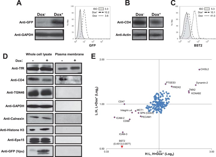
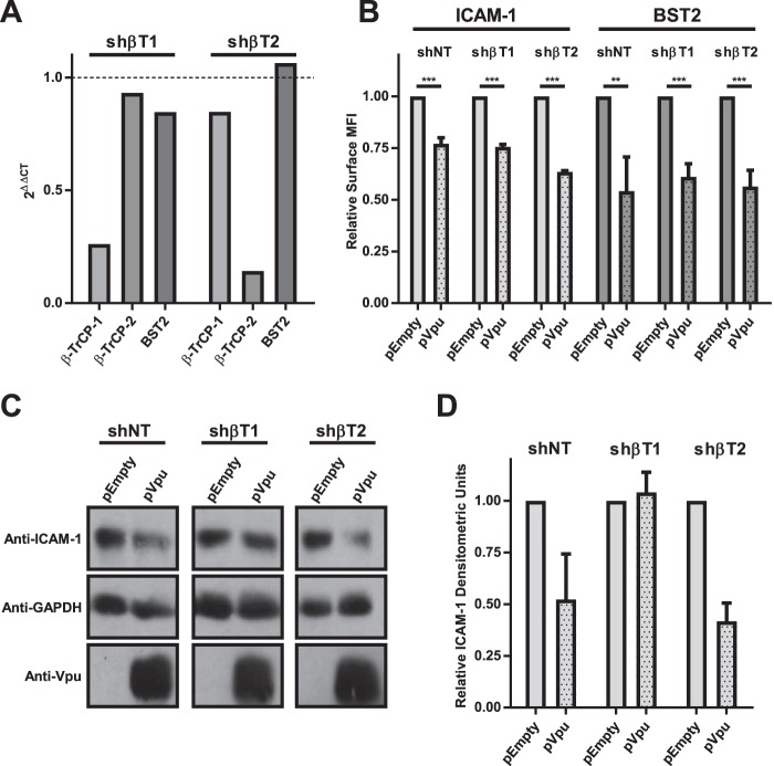
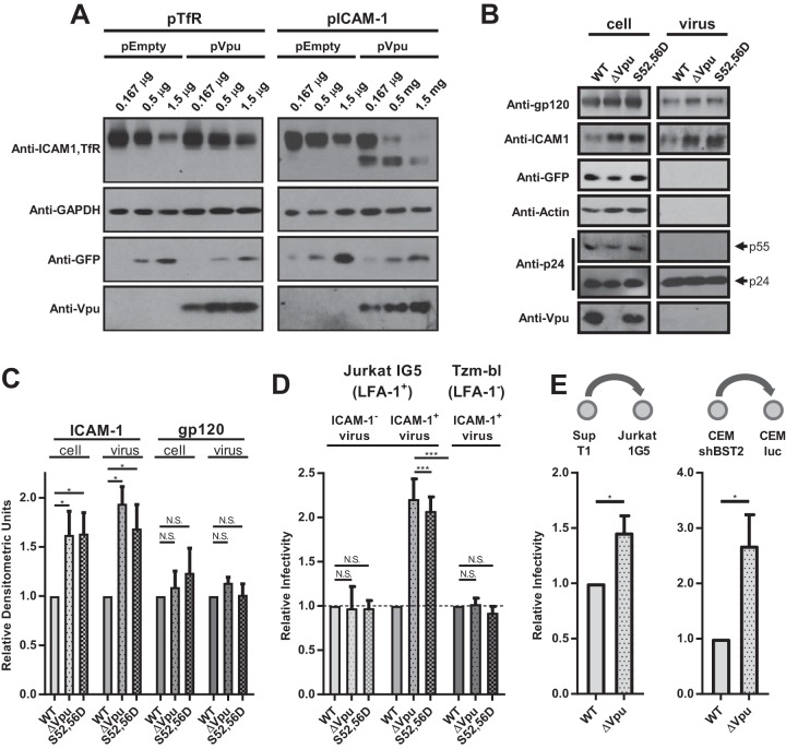
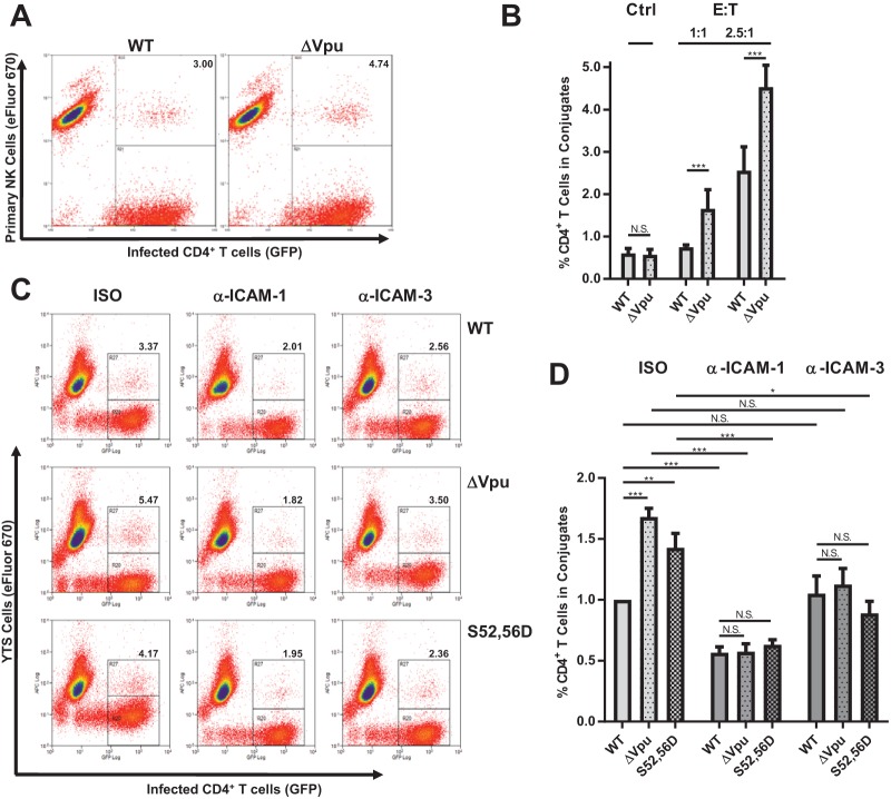
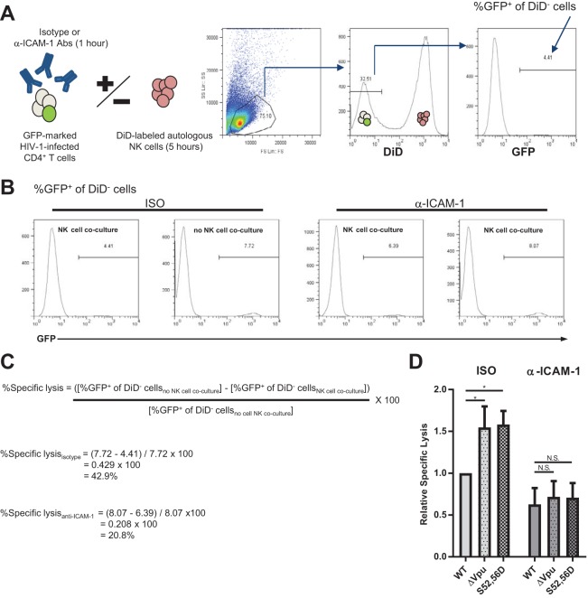
HIV-1 Vpu is known to alter the expression of numerous cell surface molecules. Given the ever-increasing list of Vpu targets identified to date, we undertook a proteomic screen to discover novel cell membrane proteins modulated by this viral protein. Plasma membrane proteome isolates from Vpu-inducible T cells were subjected to stable isotope labeling of amino acids in cell culture (SILAC)-based mass spectrometry analysis, and putative targets were validated by infection of primary CD4+ T cells. We report here that while intercellular adhesion molecule 1 (ICAM-1) and ICAM-3 are upregulated by HIV-1 infection, expression of Vpu offsets this increase by downregulating these molecules from the cell surface. Specifically, we show that Vpu is sufficient to downregulate and deplete ICAM-1 in a manner requiring the Vpu transmembrane domain and a dual-serine (S52/S56) motif necessary for recruitment of the beta-transducin repeat-containing E3 ubiquitin protein ligase (β-TrCP) component of the Skp, Cullin, F-box (SCFβ-TrCP) E3 ubiquitin ligase. Vpu interacts with ICAM-1 to induce its proteasomal degradation. Interestingly, the E3 ubiquitin ligase component β-TrCP-1 is dispensable for ICAM-1 surface downregulation yet is necessary for ICAM-1 degradation. Functionally, Vpu-mediated ICAM-1 downregulation lowers packaging of this adhesion molecule into virions, resulting in decreased infectivity. Importantly, while Vpu-mediated downregulation of ICAM-3 has a limited effect on the conjugation of NK cells to HIV-1-infected CD4+ T cells, downregulation of ICAM-1 by Vpu results in a reduced ability of NK cells to bind and kill infected T cells. Vpu-mediated ICAM-1 downregulation may therefore represent an evolutionary compromise in viral fitness by impeding the formation of cell-to-cell contacts between immune cells and infected T cells at the cost of decreased virion infectivity.IMPORTANCE The major barrier to eradicating HIV-1 infection is the establishment of treatment-resistant reservoirs early in infection. Vpu-mediated ICAM-1 downregulation may contribute to the evasion of cell-mediated immunity during acute infection to promote viral dissemination and the development of viral reservoirs. By aiding the immune system to clear infection prior to the development of reservoirs, novel treatments designed to disrupt Vpu-mediated ICAM-1 downregulation may be beneficial during acute infection or as a prophylactic treatment.

Keywords: ICAM-1; NK cell-mediated killing; Vpu; human immunodeficiency virus; immune evasion; immunological synapse; viral infectivity.


Figures

FIG 1
FIG 1
Identification of putative membrane-associated proteins targeted by HIV-1 Vpu. (A to C) Jurkat cells expressing a VpuGFP chimeric protein under the control of a Dox-inducible promoter (JurkatTetRVpuGFP) were characterized for VpuGFP expression and activity. JurkatTetRVpuGFP cells were left untreated (Dox) or treated with Dox (Dox+) for 36 h and then analyzed for VpuGFP expression by Western blotting (A, left) and flow cytometry (A, right), for CD4 expression by Western blotting (B), and for surface BST2 expression by flow cytometry (C). (D) Plasma membranes were isolated as described in Materials and Methods. Dox+ and Dox JurkatTetRVpuGFP PM isolates and whole-cell lysates were separated by SDS-PAGE and analyzed for the indicated proteins by Western blotting. (E) Dox+/− JurkatTetRVpuGFP PM isolates were analyzed by SILAC-based MS as described in Materials and Methods. The graph shows the ratio of identified heavy-labeled proteins to light-labeled proteins (H:L) when VpuGFP was induced in heavy-labeled cells (x axis; n = 3) and the ratio of identified light-labeled proteins to heavy-labeled proteins (L:H) when VpuGFP was induced in light-labeled cells (y axis; n = 3). The coordinates of the BST2 data point exceeded the limits of the graph.
FIG 2
FIG 2
Vpu prevents ICAM-1 and ICAM-3 upregulation on the surface of primary CD4+ T cells by a process that depends on its TMD and dual-serine motif. Primary human CD4+ T cells were infected with GFP-marked NL4.3 HIV-1 either lacking Vpu (ΔVpu) or expressing wild-type Vpu (WT), Vpu lacking the dual-serine motif (S52D,56D), or Vpu lacking the TMD alanine face (A10,14,18L). Forty-eight hours postinfection, surface expression of ICAM-1, ICAM-3, CD45, and BST2 was measured on GFP+ cells by flow cytometry. (A) Representative histograms. (B) Compiled data expressed as the mean fluorescence intensity (MFI) of GFP+ cells normalized to the MFI of mock-infected controls (n ≥ 4). The error bars represent standard errors of the mean (SEM). *, P ≤ 0.05; **, P ≤ 0.01; ***, P ≤ 0.005; N.S., nonsignificant.
FIG 3
FIG 3
ICAM-1 is upregulated at the transcriptional level in response to HIV-1 infection and downregulated posttranscriptionally by Vpu. HeLa cells were infected with VSV-G-pseudotyped GFP-marked wild-type NL4.3 HIV-1 (WT) or HIV-1 lacking Vpu (ΔVpu) for 48 h (n = 3). (A) The percentage of cells expressing GFP was monitored by flow cytometry. (B) ICAM-1 and BST2 mRNA levels in infected cells were measured by RT-qPCR, with gapdh used as a reference gene. (C and D) Whole-cell lysates were analyzed for the indicated proteins by Western blotting. (C) Representative blots. (D) Compiled densitometric analysis of ICAM-1 and TfR normalized to actin and expressed relative to mock-infected controls (n = 3). The error bars represent SEM. ***, P ≤ 0.005; N.S., nonsignificant.
FIG 4
FIG 4
Vpu is sufficient to deplete endogenous ICAM-1 in a TMD- and dual-serine-motif-dependent manner. HeLa cells were transfected to express wild-type NL4.3 Vpu (pVpuWT), a mutant Vpu lacking the dual-serine motif (pVpuS52,56N), a mutant Vpu containing a randomized TMD (pVpuRD), or an empty-vector control (pEmpty) for 24 h. (A and B) Surface expression of ICAM-1 was measured by flow cytometry. (A) Representative histograms of surface ICAM-1 expression. (B) Compiled data expressed as the MFI of GFP+ cells normalized to pEmpty transfections (n ≥ 3). (C and D) Whole-cell lysates were analyzed for the indicated proteins by Western blotting. (C) Representative blots. (D) Compiled densitometric analysis of ICAM-1 and TfR blots normalized to GAPDH and expressed relative to pEmpty controls (n = 2). (E) Sequence alignment of all Vpu variants tested for ICAM-1 downregulation. Structural domains are indicated above, with NL4.3 used as a consensus sequence. The arrows indicate alanine residues involved in BST2 binding. The circles indicate serine phosphorylation sites required for β-TrCP binding. (F) Primary human CD4+ T cells were infected with GFP-marked WT HIV-1 or HIV-1 lacking Vpu (ΔVpu) from the ADA-NL4.3 chimeric virus expressing ADA Vpu from an NL4.3 backbone (ADA Vpu). Surface expression of ICAM-1 was measured by flow cytometry (n = 5). The data are expressed as the MFI of GFP+ cells relative to the MFI of mock-infected controls. (G and H) HeLa cells were transfected with GFP-marked plasmids expressing WT NL4.3 Vpu (pVpuNL4.3), Vpu variants from clade B or C HIV-1 primary isolates (pVpu-B [JR-CSF] or pVpu-C [KE.00], respectively), or an empty-vector control expressing GFP alone (pEmpty) for 24 h. (G) Surface expression of ICAM-1 and BST2 was measured by flow cytometry (n = 3). The data are expressed as the MFI of GFP+ cells relative to the MFI of GFP+ cells from pEmpty transfections. (H) Whole-cell lysates were separated by SDS-PAGE and analyzed for the indicated proteins by Western blotting. A representative of 3 experiments is shown. The error bars represent SEM. *, P ≤ 0.05; ***, P ≤ 0.005.
FIG 5
FIG 5
Vpu interacts with ICAM-1 and induces its turnover in a proteasome-dependent manner. (A) HeLa cells were infected with VSV-G-pseudotyped GFP-marked wild-type (WT); Vpu-deficient (ΔVpu); or A10/A14/A18L Vpu (A10,14,18L) NL4.3 HIV-1 for 48 h prior to cell lysis and immunoprecipitation (IP) using anti-ICAM-1 Abs. The immunoprecipitates were analyzed for the indicated proteins by Western blotting. (B and C) HeLa cells were transfected to express wild-type NL4.3 Vpu (pVpu) or an empty-vector control (pEmpty) for 22 h. The cells were then pulse-labeled with 35S-labeled amino acids for 1 h and chased for the indicated times in the presence or absence of MG132 or CMA. Whole-cell lysates were immunoprecipitated with anti-ICAM-1 Abs. (B) Representative phosphorimages. The bands of interest are indicated. DMSO, dimethyl sulfoxide. (C) Relative quantification using quantitative scanning of ICAM-1 bands. The relative amount of ICAM-1 remaining over time compared to time zero is plotted for each condition (n ≥ 3). The error bars represent SEM. *, P ≤ 0.05.
FIG 6
FIG 6
Vpu dual-serine-motif-dependent surface ICAM-1 downregulation is dissociable from β-TrCP-1-mediated ICAM-1 degradation. (A) Expression of β-TrCP-1, β-TrCP-2, and BST2 mRNA transcripts was evaluated by RT-qPCR in HeLa cells expressing nontargeting shRNA (shNT) or shRNA against either β-TrCP-1 (shβT1) or β-TrCP-2 (shβT2). Data were expressed relative to the value for the shNT control. gapdh was used as a reference gene. (B to D) shNT, shβT1, and shβT2 HeLa cells shNT, shT1, and TrCP-2 were transfected with plasmids expressing wild-type NL4.3 Vpu (pVpu) or an empty-vector control (pEmpty) for 24 h. (B) Surface expression of ICAM-1 and BST2 was measured by flow cytometry (n = 3). The data are expressed as the MFI of Vpu-transfected cells normalized to the MFI of cells from pEmpty transfections. (C and D) Whole-cell lysates were analyzed for the indicated proteins by Western blotting. (C) Representative blots. (D) Compiled densitometric analysis of ICAM-1 normalized to GAPDH (n = 2). **, P ≤ 0.01; ***, P ≤ 0.005.
FIG 7
FIG 7
Vpu lowers viral infectivity by decreasing the amount of mature ICAM-1 packaged into HIV-1 virions. (A) HEK 293T cells were cotransfected with plasmids encoding ICAM-1 (pICAM-1) or TfR (pTfR), along with GFP-marked plasmids expressing wild-type NL4.3 Vpu (pVpu) or an empty-vector control expressing GFP alone (pEmpty). Twenty-four hours posttransfection, whole-cell lysates were analyzed for the indicated proteins by Western blotting. (B and C) HEK 293T cells were cotransfected with pICAM-1, along with either WT, Vpu-deficient (ΔVpu), or S52/S56D Vpu (S52, S56D) NL4.3 provirus for 48 h. Cells and virions were then pelleted and lysed prior to analysis for the indicated proteins by Western blotting. (B) Representative blots. (C) Compiled densitometric analysis of ICAM-1 and gp120 blots. Cell lysates were normalized to the amount of Gag per cell [(p24 + p55)/actin] to control for transfection efficiency. The virus lysates were normalized to p24. The results are expressed relative to the WT-infected condition (n ≥ 3). (D) Jurkat-1G5 or HeLa Tzm-bl cells expressing luciferase under the control of the HIV-1 LTR promoter were infected for 48 h with WT, ΔVpu, or S52/S56D Vpu (S52,56D) NL4.3 viruses produced from HEK 293 T cells expressing ICAM-1 or not. Luciferase activity was calculated by measuring relative light units (RLU) from cell lysates. Relative infectivity is expressed as RLU normalized to WT-infected cells (n ≥ 3). (E) Jurkat-1G5 or CEM-luc cells expressing luciferase under the control of the HIV-1 LTR promoter were infected for 48 h with WT or ΔVpu NL4.3 viruses produced from SupT1 or CEM-shBST2 cells, respectively. Luciferase activity was calculated by measuring RLU from cell lysates. Relative infectivity is expressed as RLU normalized to WT-infected cells (n = 3). A schematic representation of producer/reporter cell pairs is shown above the corresponding graphs. The error bars represent SEM. *, P ≤ 0.05; ***, P ≤ 0.005; N.S., nonsignificant.
FIG 8
FIG 8
Vpu-induced ICAM-1 downregulation prevents NK cell conjugation to HIV-1-infected CD4+ T cells. (A and B) Sorted GFP+ WT or Vpu-deficient (ΔVpu) HIV-1-infected primary CD4+ T cells were cocultured with eFluor 670-labeled autologous NK cells at different E:T ratios, and conjugate formation was measured by flow cytometry. (A) Representative dot plots at an E:T ratio of 2.5:1. The percentages of GFP+ event that were also eFluor 670+ are shown in the upper right corners. (B) Compiled data expressed as the percentage of GFP+ eFluor 670+ events out of total GFP+ events (n ≥ 4). As a control (Ctrl), 2% PFA was added to NK cells to fix them prior to CD4+ T cell coculture. Any remaining conjugation events were assumed to be the result of a nonspecific process. (C and D) Sorted GFP+ WT, Vpu-deficient (ΔVpu), or S52/S56D Vpu (S52,56D) NL4.3 HIV-1-infected primary CD4+ T cells were cocultured with eFluor 670-labeled YTS cells at an E:T ratio of 3:1 in the presence of either ICAM-1 blocking Abs (α-ICAM-1), ICAM-3 blocking Abs (α-ICAM-3), or isotype control Abs (ISO). (C) Representative dot plots. The percentages of GFP+ events that were also eFluor 670+ are shown in the upper right corners. (D) Compiled data expressed as the percentage of GFP+ eFluor 670+ events out of total GFP+ events relative to the WT isotype Ab-treated samples (n ≥ 3). The error bars represent SEM. *, P ≤ 0.05; **, P ≤ 0.01; ***, P ≤ 0.005; N.S., nonsignificant.
FIG 9
FIG 9
Vpu-induced ICAM-1 downregulation prevents NK cell-mediated killing of HIV-1-infected CD4+ T cells. Primary CD4+ T cells infected with GFP-marked WT, Vpu-deficient (ΔVpu), or S52/S56D Vpu (S52,56D) NL4.3 HIV-1 were cocultured with DiD-labeled autologous NK cells in the presence of ICAM-1 blocking Abs (α-ICAM-1) or isotype control Abs (ISO), and the percent specific lysis of HIV-1-infected CD4+ T cells was calculated by flow cytometry. (A) Experimental design and gating strategy. HIV-1-infected CD4+ T cells were pretreated for 1 h with either isotype or anti-ICAM-1 Abs before being left untreated or being cocultured with autologous, DiD dye-marked NK cells at an E:T ratio of 3:1 for 5 h. The percent GFP expression on CD4+ T cells was then determined via flow cytometry by first gating on the DiD cell population. (B) Representative data showing percent GFP expression on CD4+ T cells with or without 5 h of NK cell coculture in the presence of isotype Abs or ICAM-1 blocking Abs. (C) For both antibody treatments, the percent specific lysis of infected CD4+ T cells was calculated by dividing the decrease in percent GFP+ CD4+ T cells resulting from NK cell coculture [(percent GFP+ of DiD cellsno NK cell coculture) − (percent GFP+ of DiD cellsNK cell coculture)] by the percent GFP+ CD4+ T cells without NK cell coculture [(percent GFP+ of DiD cellsno NK cell coculture) × 100]. (D) Compiled data expressed relative to WT-infected, ISO-treated samples (n ≥ 4). The error bars represent SEM. *, P ≤ 0.05; N.S., nonsignificant.

Similar articles

Cited by

References

    1. Cohen EA, Terwilliger EF, Sodroski JG, Haseltine WA. 1988. Identification of a protein encoded by the vpu gene of HIV-1. Nature 334:532–534. doi:10.1038/334532a0. - DOI - PubMed
    1. Strebel K, Klimkait T, Martin MA. 1988. A novel gene of HIV-1, vpu, and its 16-kilodalton product. Science 241:1221–1223. doi:10.1126/science.3261888. - DOI - PubMed
    1. Salazar-Gonzalez JF, Salazar MG, Keele BF, Learn GH, Giorgi EE, Li H, Decker JM, Wang S, Baalwa J, Kraus MH, Parrish NF, Shaw KS, Guffey MB, Bar KJ, Davis KL, Ochsenbauer-Jambor C, Kappes JC, Saag MS, Cohen MS, Mulenga J, Derdeyn CA, Allen S, Hunter E, Markowitz M, Hraber P, Perelson AS, Bhattacharya T, Haynes BF, Korber BT, Hahn BH, Shaw GM. 2009. Genetic identity, biological phenotype, and evolutionary pathways of transmitted/founder viruses in acute and early HIV-1 infection. J Exp Med 206:1273–1289. doi:10.1084/jem.20090378. - DOI - PMC - PubMed
    1. Dave VP, Hajjar F, Dieng MM, Haddad E, Cohen EA. 2013. Efficient BST2 antagonism by Vpu is critical for early HIV-1 dissemination in humanized mice. Retrovirology 10:128. doi:10.1186/1742-4690-10-128. - DOI - PMC - PubMed
    1. Sato K, Misawa N, Fukuhara M, Iwami S, An DS, Ito M, Koyanagi Y. 2012. Vpu augments the initial burst phase of HIV-1 propagation and downregulates BST2 and CD4 in humanized mice. J Virol 86:5000–5013. doi:10.1128/JVI.07062-11. - DOI - PMC - PubMed

Publication types

MeSH terms

Substances

Grants and funding

LinkOut - more resources