. 2017 Mar 29;91(8):e02309-16.
doi: 10.1128/JVI.02309-16. Print 2017 Apr 15.

Computational Prediction of the Heterodimeric and Higher-Order Structure of gpE1/gpE2 Envelope Glycoproteins Encoded by Hepatitis C Virus

Affiliations

Computational Prediction of the Heterodimeric and Higher-Order Structure of gpE1/gpE2 Envelope Glycoproteins Encoded by Hepatitis C Virus

Holly Freedman et al. J Virol. .

Abstract

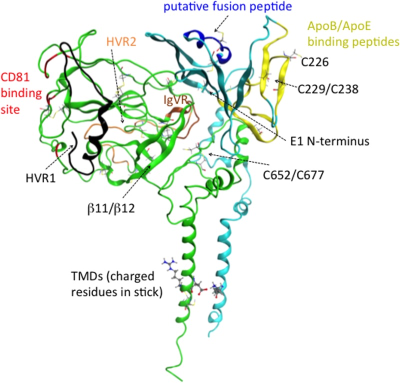
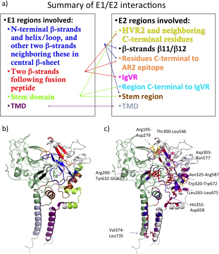
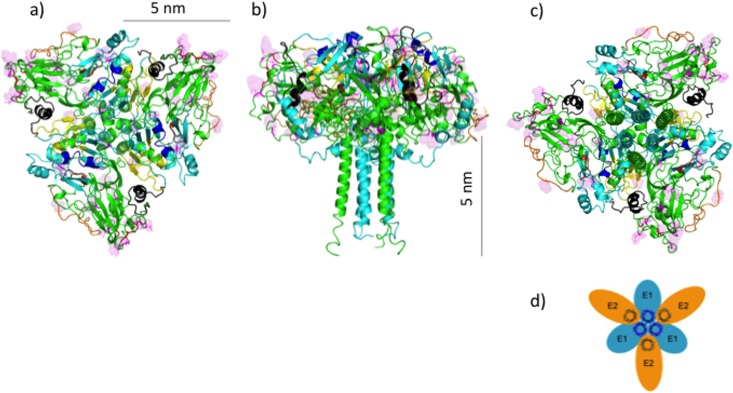
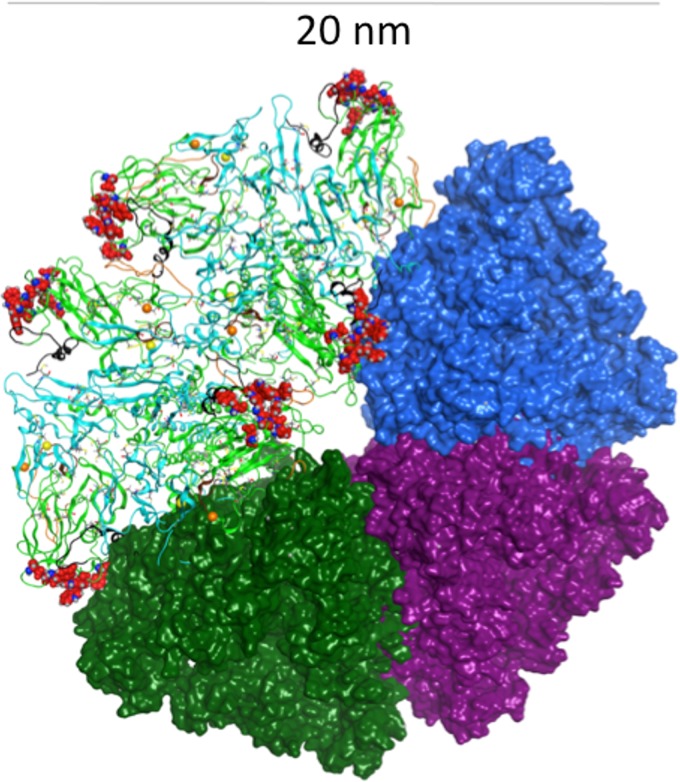
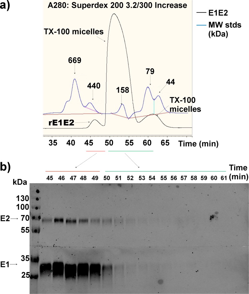
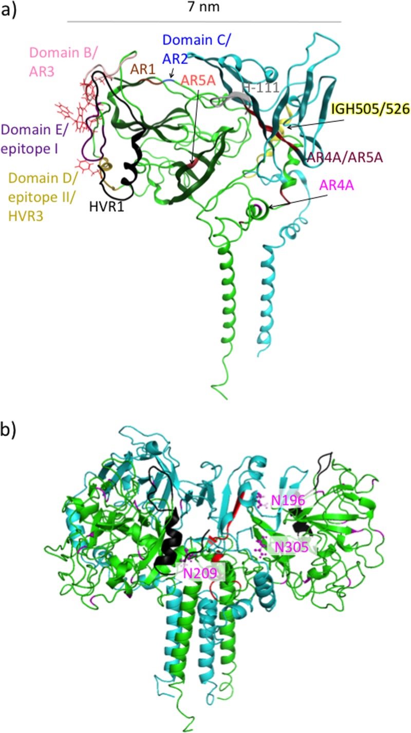
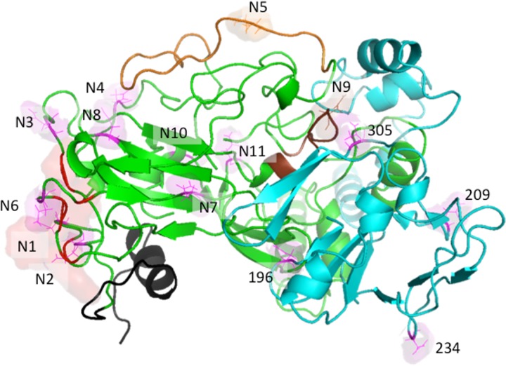
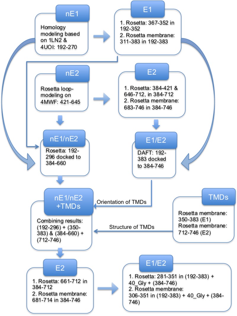
Despite the recent success of newly developed direct-acting antivirals against hepatitis C, the disease continues to be a global health threat due to the lack of diagnosis of most carriers and the high cost of treatment. The heterodimer formed by glycoproteins E1 and E2 within the hepatitis C virus (HCV) lipid envelope is a potential vaccine candidate and antiviral target. While the structure of E1/E2 has not yet been resolved, partial crystal structures of the E1 and E2 ectodomains have been determined. The unresolved parts of the structure are within the realm of what can be modeled with current computational modeling tools. Furthermore, a variety of additional experimental data is available to support computational predictions of E1/E2 structure, such as data from antibody binding studies, cryo-electron microscopy (cryo-EM), mutational analyses, peptide binding analysis, linker-scanning mutagenesis, and nuclear magnetic resonance (NMR) studies. In accordance with these rich experimental data, we have built an in silico model of the full-length E1/E2 heterodimer. Our model supports that E1/E2 assembles into a trimer, which was previously suggested from a study by Falson and coworkers (P. Falson, B. Bartosch, K. Alsaleh, B. A. Tews, A. Loquet, Y. Ciczora, L. Riva, C. Montigny, C. Montpellier, G. Duverlie, E. I. Pecheur, M. le Maire, F. L. Cosset, J. Dubuisson, and F. Penin, J. Virol. 89:10333-10346, 2015, https://doi.org/10.1128/JVI.00991-15). Size exclusion chromatography and Western blotting data obtained by using purified recombinant E1/E2 support our hypothesis. Our model suggests that during virus assembly, the trimer of E1/E2 may be further assembled into a pentamer, with 12 pentamers comprising a single HCV virion. We anticipate that this new model will provide a useful framework for HCV envelope structure and the development of antiviral strategies.IMPORTANCE One hundred fifty million people have been estimated to be infected with hepatitis C virus, and many more are at risk for infection. A better understanding of the structure of the HCV envelope, which is responsible for attachment and fusion, could aid in the development of a vaccine and/or new treatments for this disease. We draw upon computational techniques to predict a full-length model of the E1/E2 heterodimer based on the partial crystal structures of the envelope glycoproteins E1 and E2. E1/E2 has been widely studied experimentally, and this provides valuable data, which has assisted us in our modeling. Our proposed structure is used to suggest the organization of the HCV envelope. We also present new experimental data from size exclusion chromatography that support our computational prediction of a trimeric oligomeric state of E1/E2.

Keywords: E1/E2 heterodimer; Martini force field; Rosetta program; computational structural biology; hepatitis C virus.


Figures

FIG 1
FIG 1
Ribbon representation of our computational model of the E1/E2 heterodimer, with E1 in teal and E2 in green and with some of the regions discussed in the text highlighted by using color and labeling. Residues important for CD81 binding are shown in red, HVR1 is in black, HVR2 is in orange, IgVR is in brown, the ApoB/ApoE binding peptides are in yellow, and the putative fusion peptide is in dark blue. All cysteine residues are shown in a line representation.
FIG 2
FIG 2
(a and b) Structural alignment of our nE1 homology model (yellow) with structures reported under PDB accession numbers 1LN2 (34) (dark blue) (a) and 4UOI (20) (monomers in teal and pink, respectively) (b). The α-helix and two more β-strands that were added to the C-terminal end of nE1 by Rosetta are shown in magenta and green, respectively, and a similarly positioned α-helix in the structure reported under PDB accession number 1LN2 is shown in red. (c) Sequence alignment of the nE1 sequence with the sequence corresponding to the structure reported under PDB accession number 1LN2, color-coded by similarity, with dark blue representing identity and red representing clashes. The thick colored line above the sequence reported under PDB accession number 1LN2 is color-coded by secondary structure, with blue representing turns, white representing loops, red representing helical regions, and yellow representing β-strands.
FIG 3
FIG 3
(a) nE1 model, with residues modeled after the structure reported under PDB accession number 4UOI (20) shown in dark blue and those based on homology to the structure reported under PDB accession number 1LN2 (34) in gray. (b) The part of our nE2 model taken from the crystal structure reported under PDB accession number 4MWF (19). (c and d) The same regions colored as described above for panels a and b are seen in the context of our full-length model of the E1/E2 heterodimer in a back view (c) and a front view (d).
FIG 4
FIG 4
Comparison of E1/E2 interfaces and relative orientations of E1 obtained by protein-protein docking in Rosetta (orange ribbon) or by coarse-grained Gromacs simulations of the two proteins converging to form a heterodimer within a membrane environment (brown and purple ribbons). The position of E2 is the same for all models, and it is shown in yellow and in a surface representation. The viewpoint shown in panel b is rotated approximately 90° from that shown in panel a.
FIG 5
FIG 5
(a and b) Interacting regions of E1 and E2 observed in our model of the heterodimer are shown schematically (a) and illustrated on our model colored as shown in panel a (b). (c) Pairs of residues identified by interface alanine scanning as making significant energetic contributions to the E1/E2 binding free energy are illustrated on our model. E1 is shown in light gray, and E2 is in pale green. Predicted E1/E2 interface residues for which in silico mutation to Ala significantly affected E1/E2 binding free energy are colored on a scale according to increased prediction score from blue to red to yellow (highest binding free energy difference).
FIG 6
FIG 6
(a to c) Ribbon representations of the predicted trimer of heterodimers formed by E1/E2, viewed from the top (a), side (b), and bottom (c) in relation to the viral membrane being positioned below the ectodomains. The putative fusion peptides and apolipoprotein binding peptides are shown in dark blue and yellow, respectively, on E1, which is mainly in teal. E2 is shown mainly in green, with CD81 binding residues in red, HVR1 in black, HVR2 in orange, and IgVR in brown. Sites of N-glycosylation are shown in line mode and in a surface representation and are in magenta. (d) Model proposed by Falson et al. in which E1/E2 is arranged as a trimer of heterodimers, with E1 components interacting with one another at the center of the trimer through their TMDs (33).
FIG 7
FIG 7
Model of the pentamer formed by the trimer of E1/E2 heterodimers, viewed from the top in relation to the viral membrane being positioned below the ectodomains. A hexamer forming part of this pentamer is shown here in a ribbon representation, with E1 shown in teal and E2 shown mainly in green, except that CD81 binding residues are in red (shown as balls), HVR1 is in black, HVR2 is in orange, and IgVR is in brown. Cysteine residues are shown in line mode, and sulfur atoms belonging to C304 and C486 are depicted as yellow and orange balls, respectively.
FIG 8
FIG 8
SEC of purified recombinant E1E2 (H77) (a) and Western blot analysis of the fractions of the collected eluate (b). TX100, Triton X-100; MW, molecular weight (in thousands).
FIG 9
FIG 9
Ribbon representations of our computational model of the E1/E2 heterodimer, with E1 shown in teal and E2 shown in green. (a) The binding epitopes of classes of neutralizing antibodies are highlighted by using color and labeling. Residues important for CD81 binding are shown in red and in a line representation. Arrows representing suggested angles of approach are shown for the AR4A and AR5A antibodies. (b) Residues important for binding by the AR4A antibody are shown highlighted in red on one (right-hand side) of two adjacent E1/E2 heterodimers structured as part of a trimer of heterodimers. Three sites of N-linked glycosylation belonging to E1, which are close to the antibody binding site, are shown in magenta and in a ball-and-stick representation.
FIG 10
FIG 10
Locations of N-linked glycosylation sites on E1/E2, shown in line and surface representations, viewed from the top in relation to the viral membrane being positioned below the ectodomains. E1 is shown in teal, and E2 is shown mainly in green, with CD81 binding residues in red (a surface representation is also shown), HVR1 in black, HVR2 in orange, and IgVR in brown. N-glycosylation sites belonging to E2 are labeled N1 to N11, while those belonging to E1 are labeled by residue numbers.
FIG 11
FIG 11
Flowchart illustrating the computational procedure used to obtain the final model of E1/E2. For Rosetta ab initio modeling or Rosetta membrane ab initio modeling, we indicate the residues being modeled and the context in which these residues were modeled with all other residues being fixed.

References

    1. Freedman H, Logan MR, Law JL, Houghton M. 2016. Structure and function of the hepatitis C virus envelope glycoproteins E1 and E2: antiviral and vaccine targets. ACS Infect Dis 2:749–762. doi: 10.1021/acsinfecdis.6b00110. - DOI - PubMed
    1. Thomssen R, Bonk S, Propfe C, Heermann KH, Kochel HG, Uy A. 1992. Association of hepatitis C virus in human sera with beta-lipoprotein. Med Microbiol Immunol 181:293–300. doi: 10.1007/BF00198849. - DOI - PubMed
    1. Thomssen R, Bonk S, Thiele A. 1993. Density heterogeneities of hepatitis C virus in human sera due to the binding of beta-lipoproteins and immunoglobulins. Med Microbiol Immunol 182:329–334. - PubMed
    1. Andre P, Komurian-Pradel F, Deforges S, Perret M, Berland JL, Sodoyer M, Pol S, Brechot C, Paranhos-Baccala G, Lotteau V. 2002. Characterization of low- and very-low-density hepatitis C virus RNA-containing particles. J Virol 76:6919–6928. doi: 10.1128/JVI.76.14.6919-6928.2002. - DOI - PMC - PubMed
    1. Voisset C, Dubuisson J. 2004. Functional hepatitis C virus envelope glycoproteins. Biol Cell 96:413–420. doi: 10.1016/j.biolcel.2004.03.008. - DOI - PubMed

Publication types

Substances

LinkOut - more resources