. 2017 Feb 1;9(375):eaah3438.
doi: 10.1126/scitranslmed.aah3438.

Therapy with CTLA4-Ig and an antiviral monoclonal antibody controls chikungunya virus arthritis

Affiliations

Therapy with CTLA4-Ig and an antiviral monoclonal antibody controls chikungunya virus arthritis

Jonathan J Miner et al. Sci Transl Med. .

Abstract

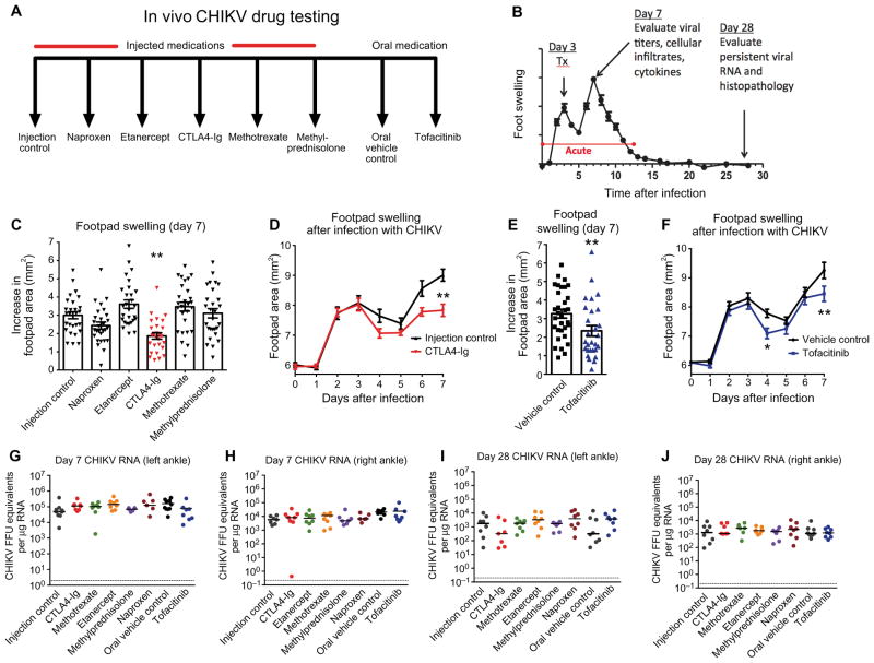
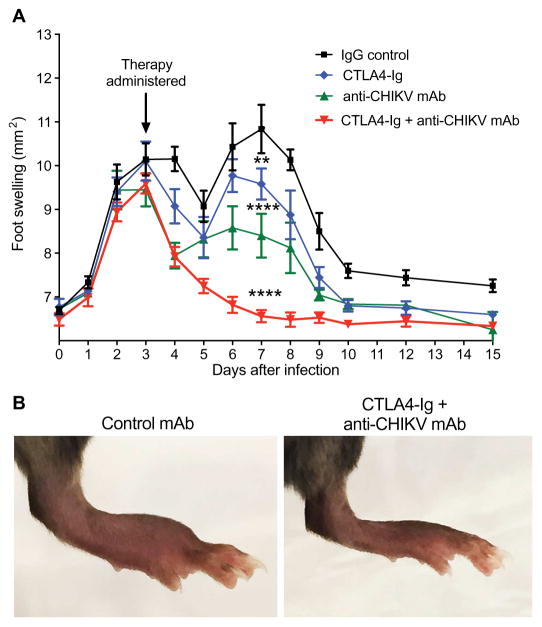
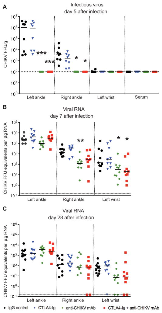
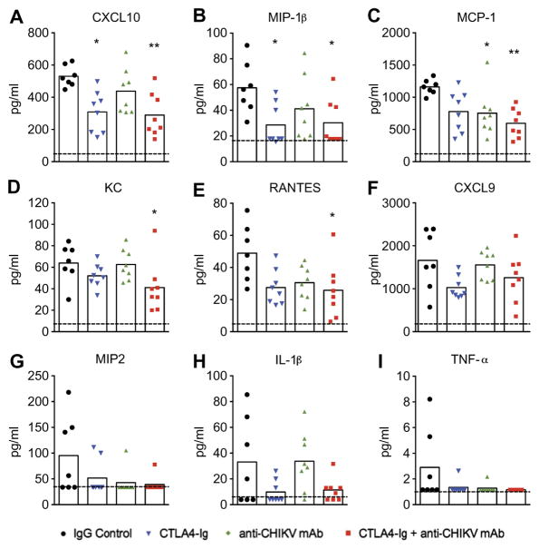
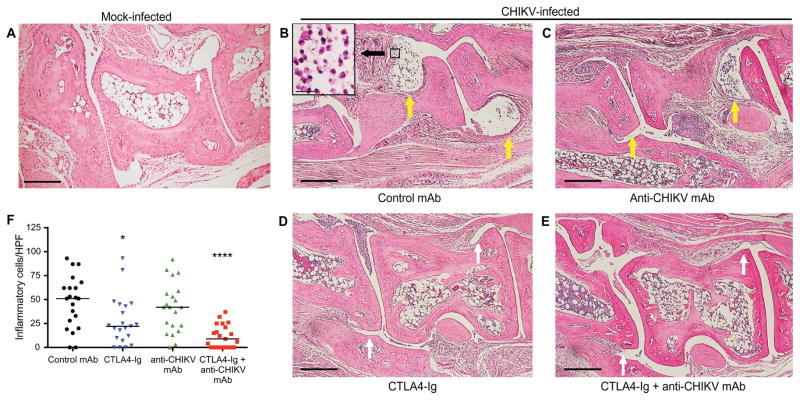
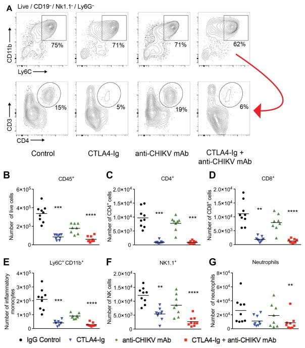
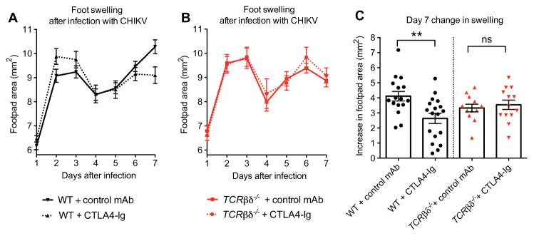
In 2013, chikungunya virus (CHIKV) transmission was documented in the Western Hemisphere, and the virus has since spread throughout the Americas with more than 1.8 million people infected in more than 40 countries. CHIKV targets the joints, resulting in symmetric polyarthritis that clinically mimics rheumatoid arthritis and can endure for months to years. At present, no approved treatment is effective in preventing or controlling CHIKV infection or disease. We treated mice with eight different disease-modifying antirheumatic drugs and identified CLTA4-Ig (abatacept) and tofacitinib as candidate therapies based on their ability to decrease acute joint swelling. CTLA4-Ig reduced T cell accumulation in the joints of infected animals without affecting viral infection. Whereas monotherapy with CTLA4-Ig or a neutralizing anti-CHIKV human monoclonal antibody provided partial clinical improvement, therapy with both abolished swelling and markedly reduced levels of chemokines, proinflammatory cytokines, and infiltrating leukocytes. Thus, combination CTLA4-Ig and antiviral antibody therapy controls acute CHIKV infection and arthritis and may be a candidate for testing in humans.


Conflict of interest statement

Competing interests: M.S.D. is a consultant for InBios, Visterra, Sanofi, and Takeda Pharmaceuticals; is on the Scientific Advisory Boards of Moderna and OraGene; and is a recipient of research grants from Moderna, Sanofi, and Visterra. J.E.C. is a consultant for Sanofi; is on the Scientific Advisory Boards of PaxVax, CompuVax, GigaGen, Meissa Vaccines, and Rensavir; is a co-inventor of the 4N12 antibody for which a patent application has been filed; and is a recipient of research grants from Moderna and Sanofi. All other authors declare that they have no competing interests.

Figures

Fig. 1
Fig. 1. Screen of candidate oral and biological DMARDs for treatment of acute CHIKV arthritis in mice
Mice were inoculated with 103 FFU of CHIKV (La Reunion strain) via a subcutaneous route. (A) Schematic depicting the two controls [phosphate-buffered saline (PBS) injection control and oral vehicle control] and six treatment arms in our drug screen. Medications and doses were methylprednisolone (0.5 mg/kg) via intraperitoneal route daily from days 3 through 7, naproxen (10 mg/kg, intraperitoneally) daily from day 3 through 7, methotrexate (0.3 mg/kg, intraperitoneally) once weekly (day 3 and day 10) 2× doses, etanercept (300 μg, intraperitoneally) 1× dose on day 3, CTLA4-Ig (300 μg, intraperitoneally) 1× dose on day 3, and tofacitinib (50 mg/kg) oral gavage daily from day 3 through day 7. For subsequent studies of CTLA4-Ig, an isotype control antibody was used. (B) Experimental design included treatment on day 3 after infection and harvests on days 7 and 28 after infection for the indicated analyses including viral burden, histology, cytokine analysis, and flow cytometry of infiltrating leukocytes. Throughout the time course, foot swelling was measured using digital calipers. (C) Foot swelling (area in square millimeter) on day 7 compared to day 0 for all injected therapies. (D) Foot swelling over time for injection control and CTLA4-Ig–treated animals. (E) Foot swelling on day 7 compared to day 0 for tofacitinib and oral vehicle control. (F) Foot swelling over time for oral vehicle control and tofacitinib-treated animals. (G to J) Real-time quantitative reverse transcription polymerase chain reaction (qRT-PCR) analysis of viral RNA levels in the left and right ankles at days 7 and 28 after infection for all treatment groups. Results in (C) to (J) are from at least two independent experiments with n = 28 per treatment group for measurement data from days 0 to 7 and n = 7 or 8 for viral burden analysis on days 7 and 28. Data represent the means (C to F) or median (G to J) ± SEM. *P < 0.05, **P < 0.005, ****P < 0.0001 [two-way analysis of variance (ANOVA) for analysis of swelling curves, Mann-Whitney for day 7 tofacitinib analysis, and Kruskal-Wallis with Dunn’s post hoc analysis for day 7 injected medication and viral burden analysis].
Fig. 2
Fig. 2. Combination therapy with CTLA4-Ig and an anti-CHIKV human mAb ameliorates acute CHIKV arthritis in mice
Mice were inoculated with 103 FFU of CHIKV via a subcutaneous route. (A) Foot swelling (area in square millimeter) from day 0 through day 15 in mice receiving at day 3 a single intraperitoneal injection of 600 μg of isotype control antibody, 300 μg of CTLA4-Ig, 300 μg of anti-CHIKV mAb (4N12), or a combination of 300 μg of CTLA4-Ig and 300 μg of anti-CHIKV mAb. Data are pooled from two independent experiments (n = 15 to 19 animals per group). (B) Representative photographs of ipsilateral foot swelling in the control mAb or combination therapy (CTLA4-Ig + anti-CHIKV mAb) groups. Data represent the means ± SEM. **P < 0.005, ****P < 0.0001 (two-way ANOVA with multiple comparisons).
Fig. 3
Fig. 3. Viral burden in CHIKV-infected mice treated with CTLA4-Ig and anti-CHIKV mAb
Mice were inoculated with 103 FFU of CHIKV via a subcutaneous route. Mice received at day 3 a single intraperitoneal injection of 600 μg of isotype control antibody, 300 μg of CTLA4-Ig, 300 μg of anti-CHIKV mAb, or a combination of 300 μg of CTLA4-Ig and 300 μg of anti-CHIKV mAb. (A) Infectious virus in joints and serum quantitated by focus-forming assay on day 5 after infection. (B and C) Real-time qRT-PCR analysis of viral RNA levels in the ipsilateral (left) and contralateral (right) ankles and left wrist at days 7 and 28 after infection. Results are pooled from two independent experiments with n = 10 per treatment group for viral RNA data and n = 8 for focus-forming assay data. Data represent the median ± SEM. *P < 0.05, **P < 0.005, ***P < 0.0005 (Kruskal-Wallis with Dunn’s post hoc analysis).
Fig. 4
Fig. 4. Cytokine and chemokine analysis in CHIKV-infected mice treated with CTLA4-Ig and anti-CHIKV mAb
Mice were inoculated with 103 FFU of CHIKV via a subcutaneous route. Mice received at day 3 a single intraperitoneal injection of 600 μg of isotype control antibody, 300 μg of CTLA4-Ig, 300 μg of anti-CHIKV mAb, or a combination of 300 μg of CTLA4-Ig and 300 μg of anti-CHIKV mAb. (A to I) Cytokine and chemokine levels on day 7 in the ipsilateral foot of mice in each treatment group. Analytes included CXCL10 (A), MIP-1β (B), MCP-1 (C), KC (D), RANTES (E), CXCL9 (F), MIP2 (G), IL-1β (H), and TNF-α (I). Results are pooled from two independent experiments with n = 8 mice per treatment group. Data represent the means ± SEM. *P < 0.05, **P < 0.005 (Kruskal-Wallis with Dunn’s post hoc analysis).
Fig. 5
Fig. 5. Representative H&E staining of the ipsilateral midfoot joints in CHIKV-infected mice treated with CTLA4-Ig and anti-CHIKV mAb
(A to F) Mice were inoculated with either PBS (A; mock) or 103 FFU of CHIKV (B to F) via a subcutaneous route. Animals were sacrificed, and histology of the ipsilateral foot was performed on day 7 after infection. CHIKV-infected mice received at day 3 a single intraperitoneal injection of 600 μg of isotype control antibody (B), 300 μg of anti-CHIKV mAb (C), 300 μg of CTLA4-Ig (D), or a combination of 300 μg of CTLA4-Ig and 300 μg of anti-CHIKV mAb (E). The number of inflammatory cells per HPF in the midfoot synovial space was quantitated in a blinded fashion (F). Results are representative of at least two independent experiments with n = 4 per treatment group and two sections assessed per foot. Scale bars, 200 μm. Yellow arrows, moderate to severe synovitis; white arrows, absent or mild synovitis *P < 0.05, ****P < 0.0001 (Kruskal-Wallis with Dunn’s post hoc analysis).
Fig. 6
Fig. 6. Flow cytometry analysis of infiltrating leukocytes in the feet of mice on day 7 after infection with CHIKV
Mice were inoculated with 103 FFU of CHIKV via a subcutaneous route. Mice received at day 3 a single intraperitoneal injection of 600 μg of isotype control antibody, 300 μg of CTLA4-Ig, 300 μg of anti-CHIKV mAb, or 300 μg of CTLA4-Ig and 300 μg of anti-CHIKV mAb. (A) Gating strategy showing subpopulations of live CD45+ cells, including the percentages of CD11b+Ly6C+ inflammatory monocytes, followed by CD3 and CD4 expression (lower panel) in the remaining Ly6C-negative cells (red arrow) isolated from the feet of mice from each treatment group. (B to G) Total number of isolated CD45+, CD4+, CD8+, Ly6C+CD11b+, NK1.1+, and Ly6G+ cells from the feet of CHIKV-infected mice in each treatment group. Results are pooled from two independent experiments with 4 to 5 mice per group in each experiment. Data represent the means ± SEM. **P < 0.005, ***P < 0.0005, ****P < 0.0001 (Kruskal-Wallis with Dunn’s post hoc analysis).
Fig. 7
Fig. 7. Clinical assessment of the response to CTLA4-Ig treatment in CHIKV-infected WT and TCR
βδ−/− mice. Mice were inoculated with 103 FFU of CHIKV via a subcutaneous route. (A and B) Foot swelling in WT (A) and TCRβδ−/− (B) mice from day 1 through 7 in mice receiving at day 3 a single intraperitoneal injection of 300 μg of isotype control antibody or 300 μg of CTLA4-Ig. (C) Increase in foot swelling (area in square millimeter) on day 7 after infection in control- and CTLA4-Ig–treated WT and TCRβδ−/− mice. ns, not significant. Measurements were conducted in a blinded fashion. Results are pooled from two or three independent experiments with total n = 16 to 18 per group for WT animals and n = 12 to 13 per group for TCRβδ−/− animals. Data represent the means ± SEM. **P < 0.005 (two-way ANOVA with multiple comparisons for swelling curves; Mann-Whitney test for day 7 swelling).

Comment in

References

    1. Ross RW. The Newala epidemic: III. The virus: Isolation, pathogenic properties and relationship to the epidemic. J Hyg. 1956;54:177–191. - PMC - PubMed
    1. Petersen LR, Powers AM. Chikungunya: Epidemiology. F1000Res. 2016;5:F1000. - PMC - PubMed
    1. Miner JJ, Aw Yeang HX, Fox JM, Taffner S, Malkova ON, Oh ST, Kim AHJ, Diamond MS, Lenschow DJ, Yokoyama WM. Brief report: Chikungunya viral arthritis in the United States: A mimic of seronegative rheumatoid arthritis. Arthritis Rheumatol. 2015;67:1214–1220. - PMC - PubMed
    1. Morrison TE, Oko L, Montgomery SA, Whitmore AC, Lotstein AR, Gunn BM, Elmore SA, Heise MT. A mouse model of chikungunya virus–induced musculoskeletal inflammatory disease: Evidence of arthritis, tenosynovitis, myositis, and persistence. Am J Pathol. 2011;178:32–40. - PMC - PubMed
    1. Rodriguez-Morales AJ, Gil-Restrepo AF, Ramírez-Jaramillo V, Montoya-Arias CP, Acevedo-Mendoza WF, Bedoya-Arias JE, Chica-Quintero LA, Murillo-García DR, García-Robledo JE, Castrillón-Spitia JD, Londoño JJ, Bedoya-Rendón HD, Cárdenas-Pérez JJ, Cardona-Ospina JA, Lagos-Grisales GJ. Post-chikungunya chronic inflammatory rheumatism: Results from a retrospective follow-up study of 283 adult and child cases in La Virginia, Risaralda, Colombia. F1000Res. 2016;5:360. - PMC - PubMed

Publication types