. 2017 Feb 2:7:41490.
doi: 10.1038/srep41490.

The E3 ligase Mule protects the heart against oxidative stress and mitochondrial dysfunction through Myc-dependent inactivation of Pgc-1α and Pink1

Affiliations

The E3 ligase Mule protects the heart against oxidative stress and mitochondrial dysfunction through Myc-dependent inactivation of Pgc-1α and Pink1

Keith Dadson et al. Sci Rep. .

Abstract

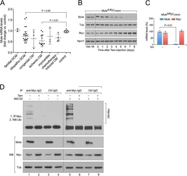
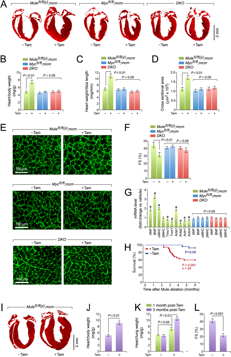
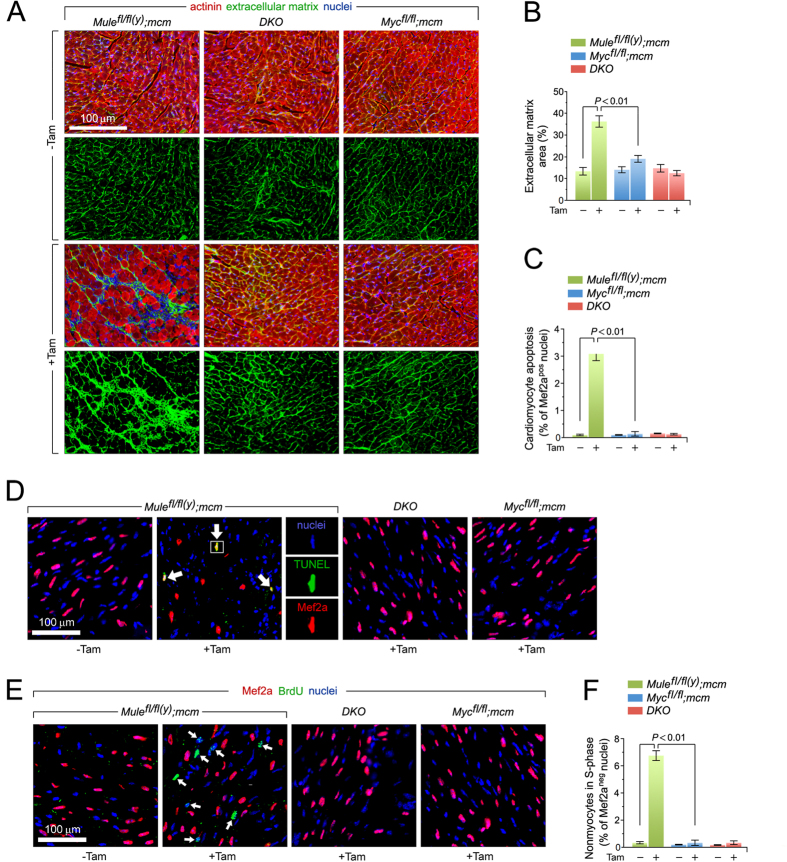
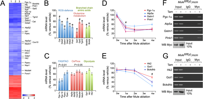
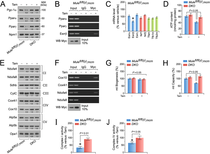
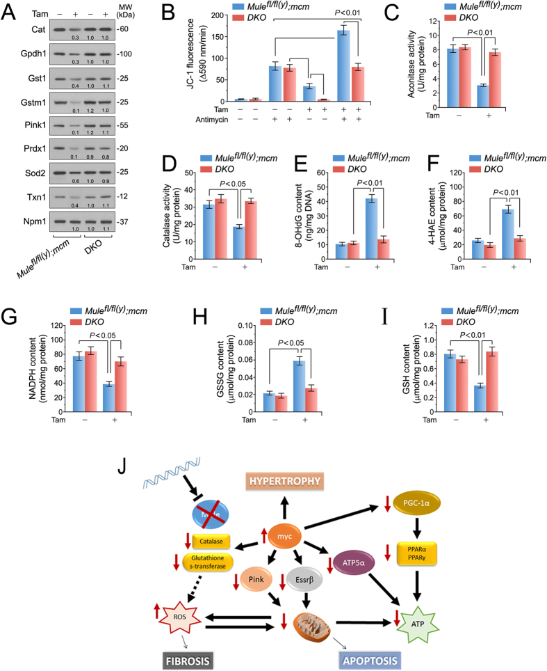
Cardiac homeostasis requires proper control of protein turnover. Protein degradation is principally controlled by the Ubiquitin-Proteasome System. Mule is an E3 ubiquitin ligase that regulates cellular growth, DNA repair and apoptosis to maintain normal tissue architecture. However, Mule's function in the heart has yet to be described. In a screen, we found reduced Mule expression in left ventricular samples from end-stage heart failure patients. Consequently, we generated conditional cardiac-specific Mule knockout (Mule fl/fl(y);mcm) mice. Mule ablation in adult Mule fl/fl(y);mcm mice prevented myocardial c-Myc polyubiquitination, leading to c-Myc accumulation and subsequent reduced expression of Pgc-1α, Pink1, and mitochondrial complex proteins. Furthermore, these mice developed spontaneous cardiac hypertrophy, left ventricular dysfunction, and early mortality. Co-deletion of Mule and c-Myc rescued this phenotype. Our data supports an indispensable role for Mule in cardiac homeostasis through the regulation of mitochondrial function via maintenance of Pgc-1α and Pink1 expression and persistent negative regulation of c-Myc.


Conflict of interest statement

The authors declare no competing financial interests.

Figures

Figure 1
Figure 1. Acute genetic deletion of Mule induces concentric hypertrophy with cardiac dysfunction and premature death.
(A) Down-regulation of Mule mRNA levels in human end-stage heart failure determined by RT- qPCR. (B) To specifically inactivate Mule and Myc in the adult heart, we employed a tamoxifen (Tam) inducible Cre-loxP system in which Cre recombinase expression is controlled by the cardiomyocyte-specific myosin heavy chain 6 promoter (mcm). 4-hydroxytamoxifen (Tam) was injected intraperitoneally daily for four consecutive days in 10 week-old mice. The day of the last Tam injection was set as day zero. Veh, vehicle. Nt, no treatment. Immunoblot analysis of cytoplasmic Mule and nuclear Myc levels in left ventricular extracts (60 μg total protein/lane) of Mulefl/fl(y);mcm mice at 7 days post-Tam employing specific antibodies as indicated on the left. Animals were 12 weeks old at the time of analysis. For normalization, Western blots were probed with anti-tubulin (Tub) for cytoplasmic fractions, and anti-nucleophosmin (Npm1) for nuclear fractions. Immunoblots were repeated at least once with similar results. Transcript levels of Mule and Myc in Mulefl/fl(y);mcm mice at 7 d post-Tam as analyzed by RT-qPCR. n = 4. (C) Mule is indispensable for Myc protein stability in the heart by regulation of its ubiquitin-mediated proteasomal degradation. At 7 d post-Tam. Mulefl/fl(y);mcm, mice were intraperitoneally injected with the proteasomal inhibitor MG132 or vehicle for 6 hours. LV lysates were immunoprecipitated (IP) with anti-Myc antibodies or normal rabbit IgG. Ubiquitinated Myc proteins in the immunoprecipitates were identified by immunoblotting with antibodies to ubiquitin. IgG, immunoglobulin G. IP, immunoprecipitation. Ubi, ubiquitin. WB, Western blot. One representative result of three independent experiments is depicted. Data are means ± s.e.m.
Figure 2
Figure 2. Genetic co-ablation of Myc and Mule prevents the cardiomyopathy associated with Mule-deficiency.
(A) Representative masson staining of longitudinal cardiac sections of the indicated mice at 4 weeks post-Tam. (B) Heart-weight corrected for body weight of the indicated strains at 4 weeks post-Tam. Animals were 15 weeks old at the time of analysis. n = 28. (C) Heart-weight corrected for tibia length at 4 weeks post-Tam. n = 28. (D) Quantification of cross-sectional area of adult cardiomyocytes at 4 weeks post-Tam. n = 12. (E) Immunofluorescence microscopy of wheat germ agglutinin (WGA; green) stained formalin-fixed LV sections at 4 weeks post-Tam. (F) Fractional shortening (FS) determined by M-mode echocardiography at 4 weeks post-Tam. n = 4. (G) Expression levels of hypertrophic and sarcomeric marker genes atrial natriuretic factor (ANP), brain natriuretic factor (BNP), α-myosin heavy chain (β-MHC), β-myosin heavy chain (β-MHC), troponin C (Tnnc1), troponin T (Tnnt1), α-actin (Acta1/2) and α-actinin (Actn1/4) as analyzed by RT-qPCR at 4 weeks post-Tam. n = 4. *P < 0.05 versus −Tam. #P < 0.01 versus −Tam. (H) Acute genetic ablation of Mule evokes premature death. Kaplan-Meier survival curves of conditional Mulefl/fl(y);mcm mice. (I) Representative masson staining of longitudinal cardiac sections from Mulefl/fl(y);mcm mice at 3 months post-Tam. (J) Heart-weight corrected for body weight of Mulefl/fl(y);mcm mice at 3 months post-Tam. n = 14. (K) Lung-weight corrected for tibia length of Mulefl/fl(y);mcm mice at the indicated time points post-Tam. n = 14. (L) Fractional shortening (FS) determined by M-mode echocardiography of Mulefl/fl(y);mcm mice at 3 months post-Tam. n = 4. Data are means ± s.e.m.
Figure 3
Figure 3. Mule-deficiency induces cardiomyocyte apoptosis and structural remodeling of the ventricular wall.
(A) Analysis of cardiac fibrosis by immunofluorescence microscopy employing wheat germ agglutinin (WGA) staining (green) of collagen deposition in the extracellular matrix, cardiomyocyte-specific anti-actinin (red), and Dapi (blue) to visualize nuclear DNA. (B) Quantification of extracellular matrix area indicative of LV fibrosis shown in (B). n = 4. (C) Acute genetic ablation of Mule triggers cardiomyocyte apoptosis which is abrogated by co- deletion of Mule and Myc in DKO mice. n = 4. (D) Analysis of apoptosis in LV cardiomyocytes (white arrows) by immunofluorescence microscopy and TUNEL assays. Hearts were harvested at 8 d post-Tam. Mice were 12 weeks old at the time of analysis. Red, cardiomyocyte-specific nuclear marker, anti-Mef2a. Green, TUNEL. Blue, DAPI stain of nuclear genomic DNA. TUNEL, terminal deoxynucleotidyl transferase- mediated dUTP nick-end-labeling. (E) BrdU, an indicator for DNA synthesis was injected intraperitoneally at 7 d post-Tam. Animals were sacrificed 18 hours later. Quantitative analysis of cardiomyocytes in S phase was performed by immunofluorescence microscopy of LV cardiac sections employing anti-BrdU (green) and anti-Mef2a (red) antibodies. White arrows denote BrdU-positive and Mef2a-negative non-cardiomyocytes. Blue, nuclei). BrdU, 5-Bromo-2′-deoxyuridine. (F) Genetic ablation of Mule fails to induce cell cycle entry and DNA synthesis in adult cardiomyocytes. n = 4. Data are means ± s.e.m.
Figure 4
Figure 4. Mule inhibits Myc-dependent deterioration of cardiac function by downregulation of Pgc-1α and Pink1.
Microarray-based genome-wide transcriptional profiling of LV tissue samples derived from Mulefl/fl(y);mcm mice at 4 weeks post-Tam. Animals were 15 weeks old at the time of analysis. Depicted are differentially expressed genes that were significantly enriched for a cellular metabolic gene signature in the Mule-deficient mice (column) post-Tam relative to vehicle- injected controls. Heatmap values (log2 fold expression) of this metabolic gene set are shown by color and intensity of shading. Important factors of cardiac energy metabolism and detoxification of oxygen radicals are indicated on the right. Blue, repressed. Red, induced. n = 3 biological replicates. P < 0.01. Fold change >2.0. (A) Expression levels of key genes selected from (A), involved in the regulation of branched chain amino acid metabolism, glutamine metabolism and ROS defence as analyzed by RT-qPCR at 4 weeks post-Tam. n = 4. *P < 0.05 versus −Tam. #P < 0.01 versus vehicle. (B) Expression levels of differentially enriched genes from (A), involved in the regulation of glycolysis, mitochondrial oxidative phosphorylation (OxPhos), fatty acid synthesis (FAS) and fatty acid oxidation (FAO) as analyzed by RT-qPCR at 4 weeks post-Tam. n = 4. (D,E) Time course of transcript levels of selected factors (C) participating in the regulation of important biological processes in Mulefl/fl(y);mcm mice was analyzed by RT-qPCR. n = 4. #P < 0.01 versus vehicle. (F) Endogenous Myc interacts with gene promoter sequences of Pgc-1α, Atp5a, Gstm1 and Pink1 in situ. One representative result of three independent experiments is shown. (G) Myc does not bind to the promoters of Hk2, Got1 and Bckdhb. LV tissue obtained from Mulefl/fl(y);mcm mice at 7 d post-Tam was analyzed by ChIP employing anti-Myc antibodies and PCR primers specific to the selected gene promoters. Normal rabbit IgG were used for control immunoprecipitations. Input lanes of chromatin levels used for the immunoprecipitation step demonstrate equal loading. One representative result of three independent experiments is shown. Data are means ± s.e.m.
Figure 5
Figure 5. Mule is indispensable for the maintenance of mitochondrial biogenesis and bioenergetics.
(A) Immunoblot analysis of master transcription factors regulating FAS and FAO in left ventricular extracts of Mulefl/fl(y);mcm and DKO mice at 7 days post-Tam. Numbers below the individual blots indicate fold-changes in protein expression. (B) Endogenous Myc interacts with gene promoter sequences of Esrrβ, but is not associated with the promoters of Pparα and Pparγ as analyzed by ChIP. Esrrβ, estrogen related receptor, beta. (C) Transcript expression of key factors regulating FAS/FAO, redox homeostasis and mt biogenesis in left ventricular samples of Mulefl/fl(y);mcm mice was determined by RT-qPCR at 4 weeks post-Tam. n = 4. *P < 0.05 versus vehicle. (D) ATP levels in left ventricular tissue extracts from Mulefl/fl(y);mcm mice at 7 d post-Tam. n = 4. (E) Immunoblot analysis of important constituents of the mt electron transport chain participating in oxidative phosphorylation in LV extracts of Mulefl/fl(y);mcm versus DKO mice at 7 days post-Tam. Equal loading of bands was confirmed by reprobing membranes with mt- specific anti-Opa1 antibodies. (F) Myc interacts with gene promoter sequences of OxPhos components of Ndufa4 (CI, complex 1) and Cox10 (CIV, complex 4) as analyzed by ChIP from isolated LV genomic DNA from Mulefl/fl(y);mcm mice at 7 d post-Tam. (G) Mt biogenesis, defined as relative DNA copy number of mt encoded Cytb gene normalized to the copy number of the nuclear gene Npm1, was determined by qPCR of isolated LV mt from Mulefl/fl(y);mcm and DKO mice at 7 d post-Tam. n = 4. (H) Mt capacity, defined as relative mRNA levels of the nuclear gene Ndufa8, a subunit of OxPhos complex I, normalized to Npm1 transcript expression, was determined by RT-qPCR of total RNA isolated from left ventricular tissue of the indicated wild type (−Tam) and mutant strains (+Tam). n = 4. (I) Activity of OxPhos complex I in Mulefl/fl(y);mcm and DKO mice at 7 d post-Tam. n = 4. *P < 0.01 versus vehicle. (J) Activity of OxPhos complex IV in Mulefl/fl(y);mcm mice or DKO animals at 7 d post-Tam. n = 4. *P < 0.05 versus vehicle. Data are means ± s.e.m.
Figure 6
Figure 6. Enhanced oxidative stress in mice with cardiac-specific deletion of Mule.
(A) Immunoblot analysis of factors involved in detoxification processes of reactive oxygen species in left ventricular extracts of Mulefl/fl(y);mcm and DKO mice at 7 days post-Tam employing specific antibodies as indicated on the left. One representative result of three independent experiments is shown. (B) The membrane potential (ΔΨm) of mitochondria isolated from Mule mutant mice is susceptible to ROS-induced depolarization. Genetic co-ablation of Myc and Mule in DKO mice rescues antimycin induced decreases of ΔΨm in mitochondria derived from this double mutant strain. Isolated mitochondria, incubated with JC-1 (5 μg/mL), were treated with antimycin (50 μM). JC-1 emission at 535/595 nm was recorded at 1 reading/min for 30 min using a fluorescence spectrophotometer. The rate between two time points (Δemission at 595 nm/min) was calculated in the most linear range of decline for JC-1 fluorescence intensity. n = 4. (C,D) Mitochondrial aconitase (C) and catalase (D) activities were determined spectrophotometrically in ventricular samples from Mulefl/fl(y);mcm and DKO strains at 7 days post-Tam. n = 4. (E) Oxidative genomic DNA damage in the hearts of Mulefl/fl(y);mcm mice is abolished by genetic co-ablation of Myc in DKO animals. Concentrations of 8-hydroxy-2′-deoxyguanosine (8- OHdG), a biomarker for oxidative DNA damage in the indicated strains was determined by a competitive enzyme-linked ELISA employing 8-OHdG antibodies. n = 4. (F) Higher levels of 4-hydroxyalkenals (4-HAE), an indicator of ROS-dependent lipid peroxidation in ventricular extracts of Mulefl/fl(y);mcm compared with DKO. n = 4. (G) Significantly reduced levels of NADPH, an essential cofactor for the reduction of glutathione, in in ventricul*ar extracts of Mulefl/fl(y);mcm mice versus DKO. n = 4. (H,I) Decreased the reduced glutathione/oxidized glutathione (GSH/GSSG) ratios, an indicator of cardiac oxidative stress, in ventricular samples from Mulefl/fl(y);mcm mice that was prevented by Myc co-ablation in DKO. n = 4. Schematic model for Mule-mediated inhibition of Myc-dependent cardiac hypertrophy. In response to genetic ablation of Mule, Myc is activated and transcriptionally inhibits canonical downstream targets Pgc-1α and Pink1 promoting the development of heart failure by induction of oxidative stress and mitochondrial dysfunction. Data are means ± s.e.m.

Similar articles

Cited by

References

    1. Ziaeian B. & Fonarow G. C. Epidemiology and aetiology of heart failure. Nat Rev Cardiol 13, 368–378 (2016). - PMC - PubMed
    1. Jessup M. & Brozena S. Heart failure. N Engl J Med 348, 2007–2018 (2003). - PubMed
    1. Johansen H., Strauss B., Arnold J. M., Moe G. & Liu P. On the rise: The current and projected future burden of congestive heart failure hospitalization in Canada. The Canadian journal of cardiology 19, 430–435 (2003). - PubMed
    1. Rubart M. & Field L. J. Cardiac regeneration: repopulating the heart. Annu Rev Physiol 68, 29–49 (2006). - PubMed
    1. Engel F. B. et al.. p38 MAP kinase inhibition enables proliferation of adult mammalian cardiomyocytes. Genes & development 19, 1175–1187 (2005). - PMC - PubMed

MeSH terms

Substances

LinkOut - more resources