. 2017 Feb 2:7:41478.
doi: 10.1038/srep41478.

Construction of a guide-RNA for site-directed RNA mutagenesis utilising intracellular A-to-I RNA editing

Affiliations

Construction of a guide-RNA for site-directed RNA mutagenesis utilising intracellular A-to-I RNA editing

Masatora Fukuda et al. Sci Rep. .

Abstract

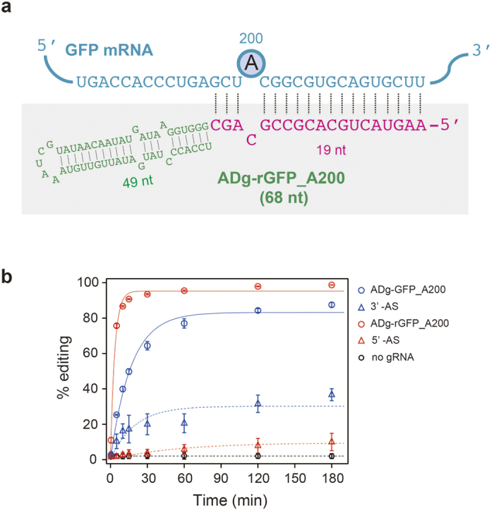
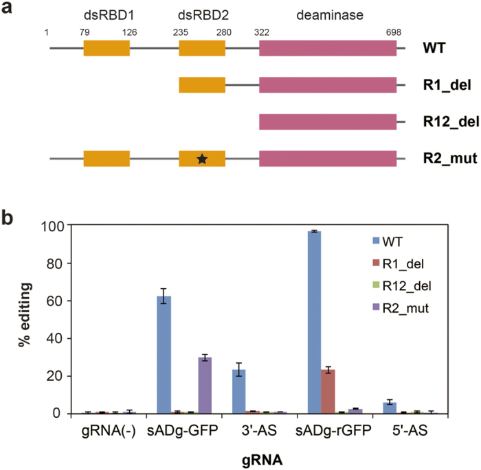
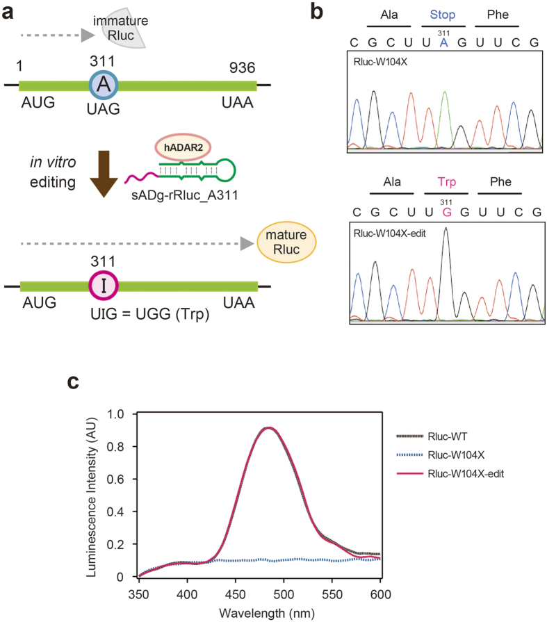
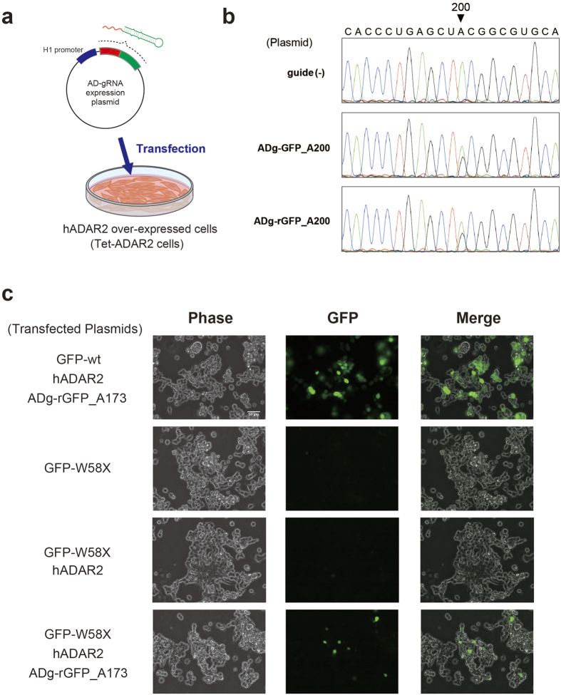
As an alternative to DNA mutagenesis, RNA mutagenesis can potentially become a powerful gene-regulation method for fundamental research and applied life sciences. Adenosine-to-inosine (A-to-I) RNA editing alters genetic information at the transcript level and is an important biological process that is commonly conserved in metazoans. Therefore, a versatile RNA-mutagenesis method can be achieved by utilising the intracellular RNA-editing mechanism. Here, we report novel guide RNAs capable of inducing A-to-I mutations by guiding the editing enzyme, human adenosine deaminase acting on RNA (ADAR). These guide RNAs successfully introduced A-to-I mutations into the target-site, which was determined by the reprogrammable antisense region. In ADAR2-over expressing cells, site-directed RNA editing could also be performed by simply introducing the guide RNA. Our guide RNA framework provides basic insights into establishing a generally applicable RNA-mutagenesis method.


Conflict of interest statement

The authors declare no competing financial interests.

Figures

Figure 1
Figure 1. Construction of gRNAs inducing hADAR2 for site-directed A-to-I RNA editing.
(a) Principle of the AD-gRNA strategy for site-directed RNA editing. Schematic representation of A-to-I RNA editing by hADAR2 is shown in the grey background. hADAR2 is composed of 2 dsRBDs and a deaminase domain. AD-gRNA, composed of an antisense region (magenta) and an ADAR-recruiting region (green) was designed based on a naturally edited substrate. A schematic representation of site-directed RNA-editing strategy using AD-gRNA is shown in the right panel. Target RNA is depicted with a turquoise line, and the black dashed line represents a base-pairing interaction. The target-editing site is marked by the circled A base. The AD-gRNA promotes both ADAR recruitment and target recognition by target-RNA hybridization. Thus, site-directed RNA editing is achieved by guiding hADAR2 onto the target site. (b) Sequence design of an AD-gRNA based on naturally edited substrate RNA. The well-known editing site in GluR2 RNA (R/G site) is indicated with the circled ‘A’. The position used to divide the GluR2 RNA into a prototype AD-guide RNA and a target-RNA is marked with a black arrowhead. The sequence of each fragment generated by the division is shown underneath. The AD-gRNA is shown within the grey background, and the antisense region (ASR) and ADAR-recruiting region (ARR) are shown in magenta and green, respectively. The target RNA is shown in turquoise characters. ‘X’ refers to any nucleotide, and the dotted lines connecting the blue and red characters indicate base pairing. (c) Editing-inducing activity of AD-gRNA. Sequencing chromatograms of the resultant sGFP cDNAs, which were obtained from RT-PCR followed by in vitro editing reaction using recombinant hADAR2 without gRNA (upper panel), with only the ASR (17 nt; middle panel), or with ADg-GFP_A200 (lower panel) are shown. Green and black peaks indicate signals for adenosine and guanosine, respectively. The target-editing site is indicated with a black arrowhead.
Figure 2
Figure 2. Design and editing induction activity of 5′-AS AD-gRNA.
(a) Nucleotide sequence of ADg-rGFP_A200 and a partial sequence of sGFP RNA are shown as a complex with a predicted secondary structure. sGFP RNA is represented in blue, and the target-editing site (A200) is depicted using a circled ‘A’. ASR and ARR are shown in red and green characters, respectively. (b) In vitro-editing induction activity of AD-gRNA. Changes in the editing ratio at A200 over time with 3′-AS AD-gRNA (ADg-GFP_A200, blue open circles), 3′-AS alone (blue open triangles), 5′-AS AD-gRNA (ADg-rGFP_A200, red open circles), 5′-AS (red open triangle), or without gRNA (black open circles). Each editing percentage was quantified by measuring the peak heights for A and G generated from the direct-sequencing chromatograms (Supplementary Figs 4 and 5) and calculated as follows: (height of the G peak)/(height of A peak + height of G peak). The results are presented as averages with standard deviations from 3 independent experiments.
Figure 3
Figure 3. Analysis of dsRBD-dependent, AD-gRNA-induced site-directed RNA editing.
(a) Schematic representation of the hADAR2 mutants used in this study. The regions corresponding to the dsRBDs and deaminase domain are represented in orange and magenta. The numbers denote the amino acid positions, relative to the N terminus of hADAR2. The black star indicates the mutation causing a defect in dsRNA binding by the dsRBD. (b) Editing percentages at A200 in sGFP RNA during the AD-gRNA editing-induction reaction, with each hADR2 mutant. Using wild-type hADAR2 (blue), R1_del (red), R12_del (green) and R2_mut (purple), editing reactions were performed without gRNA (gRNA[-]), with sADg-GFP_A200 (ADg-GFP) and its ASR (3′-AS), and with sADg-rGFP_A200 (ADg-rGFP) and its ASR (5′-AS). The editing percentages shown were calculated from the peak heights for A and G generated from the direct-sequencing chromatograms, as follows: (height of the G peak)/(height of A peak + height of G peak). The results are presented as averages with standard deviations from 3 independent experiments.
Figure 4
Figure 4. Amber codon-repair experiment by AD-gRNA-induced RNA editing.
(a) Schematic representation of a codon-repair experiment conducted using a modified luciferase reporter. The modified Renilla luciferase mRNA (Rluc-W104X) was generated by changing guanosine at nucleotide 311 to adenosine (A311) to alter codon Trp104 (UGG) into an amber stop codon (UAG). Active mature luciferase was translated from Rluc-W104X after A311 was edited to I311 by ADg-rRluc_A311. (b) Sequence chromatograms of cDNA from Rluc-W104X (upper) and in vitro-edited Rluc-W104X with ADg-rRluc_A311 (Lower). (c) Confirmation of active luciferase expression regulated by AD-gRNA. Luminescence-spectrum analysis of samples after performing an in vitro translation reaction with wild-type luciferase mRNA (Rluc-WT, black), Rluc-W104X (blue), or in vitro edited-Rluc-W104X (magenta).
Figure 5
Figure 5. Application of AD-gRNA for site-directed RNA mutagenesis and regulating target-protein expression in cells.
(a) Site-directed RNA mutagenesis with AD-gRNA following plasmid transfection in hADAR2-over-expressed cells. The expression plasmid for AD-gRNA was constructed using a pol III-driven short RNA expression vector. Previously constructed tet-ADAR2 cells, in which both hADAR2 and AcGFP expression can be controlled under a Dox-inducible promoter, were used as hADAR2-over-expressed cells. (b) Confirmation of specific editing-induction activity of ADg-GFP_A200 and ADg-rGFP_A200 in tet-ADAR2 cells. Sequencing chromatograms of GFP cDNA obtained from cells cultured without plasmid transfection (guide [-], upper), with p-ADg-GFP_A200 (middle), or p-ADg-rGFP_A200 (lower) are shown. The target adenosine (A200) is indicated with a black arrowhead. All sequencing chromatograms are shown in Supplementary Fig. 18. (c) Fluorescent microscopy pictures in the intracellular codon-repair experiment. GFP-W58X is a fluorescent reporter for real-time monitoring of functional protein expression by amber codon repair induced by AD-gRNA. The plasmids transfected are shown on the left of the pictures. The top pictures show results from control experiments, in which HEK293 cells were co-transfected with a wild-type GFP expression plasmid (GFP-wt), p-hADAR2, and p-ADg-rGFP_A173. The lower pictures show the cells co-transfected with the reporter plasmid (GFP-W58X), p-hADAR2, and p-ADg-rGFP_A173. Fluorescent micrographs for each cell were obtained at 48 h post-transfection.

References

    1. Heidenreich M. & Zhang F. Applications of CRISPR-Cas systems in neuroscience. Nat Rev Neurosci 17, 36–44 (2016). - PMC - PubMed
    1. Kim Y. G., Cha J. & Chandrasegaran S. Hybrid restriction enzymes: zinc finger fusions to Fok I cleavage domain. Proc Natl Acad Sci USA 93, 1156–1160 (1996). - PMC - PubMed
    1. Porteus M. H. & Carroll D. Gene targeting using zinc finger nucleases. Nat Biotechnol 23, 967–973 (2005). - PubMed
    1. Zhang F. et al. Efficient construction of sequence-specific TAL effectors for modulating mammalian transcription. Nat Biotechnol 29, 149–153 (2011). - PMC - PubMed
    1. Mali P. et al. RNA-guided human genome engineering via Cas9. Science 339, 823–826 (2013). - PMC - PubMed

Publication types