. 2017 Feb 2:7:41608.
doi: 10.1038/srep41608.

Palmdelphin promotes myoblast differentiation and muscle regeneration

Affiliations

Palmdelphin promotes myoblast differentiation and muscle regeneration

Yaping Nie et al. Sci Rep. .

Abstract

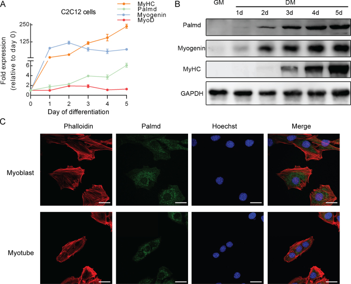
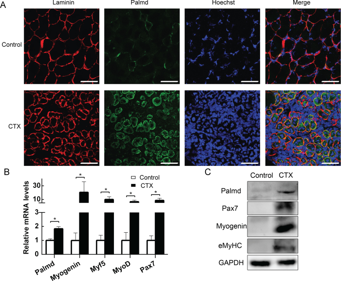
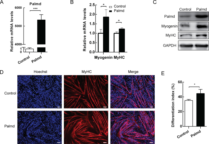
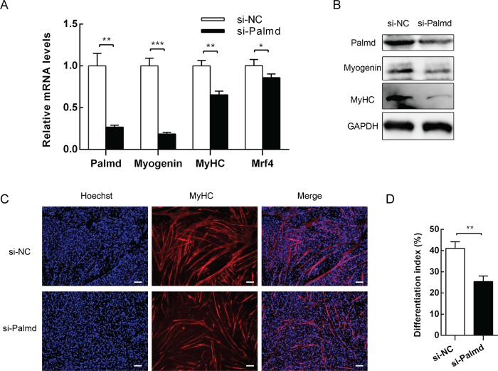
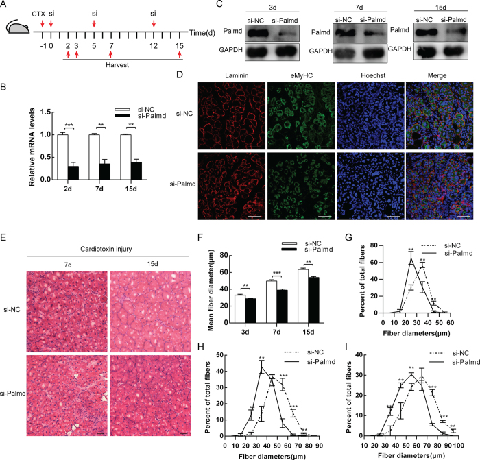
Differentiation of myoblasts is essential in the development and regeneration of skeletal muscles to form multinucleated, contractile muscle fibers. However, the process of myoblast differentiation in mammals is complicated and requires to be further investigated. In this study, we found Palmdelphin (Palmd), a cytosolic protein, promotes myoblast differentiation. Palmd is predominantly expressed in the cytosol of myoblasts and is gradually up-regulated after differentiation. Knockdown of Palmd by small interfering RNA (siRNA) in C2C12 markedly inhibits myogenic differentiation, suggesting a specific role of Palmd in the morphological changes of myoblast differentiation program. Overexpression of Palmd in C2C12 enhances myogenic differentiation. Remarkably, inhibition of Palmd results in impaired myotube formation during muscle regeneration after injury. These findings reveal a new cytosolic protein that promotes mammalian myoblast differentiation and provide new insights into the molecular regulation of muscle formation.


Conflict of interest statement

The authors declare no competing financial interests.

Figures

Figure 1
Figure 1. Palmd expression and location during C2C12 myoblast differentiation.
(A) qPCR for the Palmd, Myogenin, MyoD and MyHC expression profile during C2C12 myoblast differentiation at the indicated time points, showed up-regulation of Palmd after differentiation. Data are presented as mean ± s.e.m., n = 3 per group. (B) The protein levels of Palmd, Myogenin, MyHC during C2C12 myoblast differentiation showed a similar result. GM: growth medium, DM: differentiation medium. (C) Immunofluorescence staining for Palmd (green) together with Phalloidin (red) and nucleus (blue) staining in myoblast and myotube. Scale bar = 25 μm.
Figure 2
Figure 2. Palmd is activated in injured TA muscle.
(A) Immunofluorescence staining for Palmd (green), Laminin (red) and nucleus (blue) staining in TA muscle sections 3.5 days after CTX or saline (control) injection. Part of Palmd (green) and Laminin (red) was merged (yellow). Scale bar = 75 μm. (B) The mRNA levels of Palmd, Myogenin, Myf5, MyoD and Pax7 in TA muscles 3.5 days after CTX or saline injection. Data are presented as mean ± s.e.m, n = 3 per group. * p < 0.05. (C). The protein levels of Palmd, Myogenin, MyoD and Pax7 in TA muscles 3.5 days after CTX or saline injection.
Figure 3
Figure 3. Palmd overexpression promotes C2C12 myoblast differentiation.
C2C12 cells were transfected with pcDNA3.1 vector (control) or pcDNA3.1-Palmd vector (Palmd), then induced to differentiate 2d later. (A) The mRNA levels of Palmd 2d after differentiation in two groups above. (B) The mRNA levels of Myogenin 12 h after differentiation, and MyHC 3d after differentiation, in two groups above. (C) The protein levels of Palmd, Myogenin and MyHC at the time points described in (A) and (B). (D) After transfection and differentiation for 3 days, MyHC was detected by immunofluorescence staining. Scale bar = 100 μm. (E) The differentiation index in (D) was calculated. 3 random microscopic fields were selected for each group. Data are presented as mean ± s.e.m., n = 3 per group. *p < 0.05, ***p < 0.001.
Figure 4
Figure 4. Knockdown of Palmd attenuates C2C12 myoblast differentiation.
C2C12 cells were transfected with negative control siRNA (si-NC) pool or Palmd siRNA pool (si-Palmd), then induced to differentiate 2d later. (A) The mRNA levels of Palmd 2d after differentiation, and Myogenin 12 h after differentiation, and MyHC, Mrf4 3d after differentiation, in two groups above. (B) The protein levels of Palmd, Myogenin and MyHC at the time points described in (A). (C) After transfection and differentiation for 3 days, MyHC was detected by immunofluorescence staining. Scale bar = 100 μm. (D) The differentiation index in (C) was calculated. 3 random microscopic fields were selected for each group. Data are presented as mean ± s.e.m., n = 3 per group. *p < 0.05, **p < 0.01, ***p < 0.001.
Figure 5
Figure 5. Knockdown of Palmd in vivo impairs muscle regeneration.
(A) Schematic representation of CTX injury followed by si-NC or si-Palmd treatment in mouse TA muscle. (B) The mRNA level changes of Palmd in TA muscle at 2d, 7d and 15d, after CTX injury (−1d) and siRNA injection (0d, 5d and 12d). (C) The protein levels of Palmd in TA muscle of si-NC or si-Palmd group on 3d 7d and 15d. (D) Immunofluorescence staining for Laminin (red), eMyHC (green) and nucleus (blue) on si-NC or si-Palmd group TA muscle section on 3d. Scale bar = 75 μm. (E) H&E staining on si-NC or si-Palmd treated TA muscle section at 7d and 15d. Scale bar = 50 μm. (F) TA muscle average myofiber diameters (μm) of si-NC or si-Palmd group at 3d, 7d and 15d. n = 3 per group, more than 300 myofibers per mouse. (G~I) Percent distributions by diameter (μm) of myofibres at 3d (G), 7d (H) and 15d (I) analyzed in (F). Data are presented as mean ± s.e.m., n = 3 per group. **p < 0.01, ***p < 0.001.

Similar articles

Cited by

References

    1. Baskin K. K., Winders B. R. & Olson E. N. Muscle as a “mediator” of systemic metabolism. Cell Metab. 21, 237–48 (2015). - PMC - PubMed
    1. Apponi L. H., Corbett A. H. & Pavlath G. K. RNA-binding proteins and gene regulation in myogenesis. Trends Pharmacol Sci. 32, 652–8 (2011). - PMC - PubMed
    1. Berkes C. A. & Tapscott S. J. MyoD and the transcriptional control of myogenesis. Semin Cell Dev Biol. 16, 585–95 (2005). - PubMed
    1. Tajbakhsh S. Skeletal muscle stem and progenitor cells: reconciling genetics and lineage. Exp Cell Res. 306, 364–372 (2005). - PubMed
    1. Bentzinger C. F., Wang Y. X. & Rudnicki M. A. Building muscle: molecular regulation of myogenesis. Cold Spring Harb Perspect Biol. 1, 4(2) (2012). - PMC - PubMed

Publication types