

A Cone Beam Computed Tomography (CBCT) evaluation of MB2 canals in endodontically treated permanent maxillary molars. A retrospective study in Indian population
- PMID: 28149463
- PMCID: PMC5268108
- DOI: 10.4317/jced.52716
A Cone Beam Computed Tomography (CBCT) evaluation of MB2 canals in endodontically treated permanent maxillary molars. A retrospective study in Indian population
Abstract
Background: Current technological advances have allowed application of different study designs and techniques for investigation of dental anatomy. Some clinical studies have provided evidence that Cone Beam computed tomography (CBCT) scanning is an important resource in assessment of root canal systems notably to identify MB2 canals in maxillary molars as CBCT scans allow in vivo dental investigation in axial, sagittal and coronal planes simultaneously. The current study was undertaken to detect and evaluate filled/unfilled MB2 canals in endodontically treated, asymptomatic maxillary molars utilizing cone beam computed tomography (CBCT).
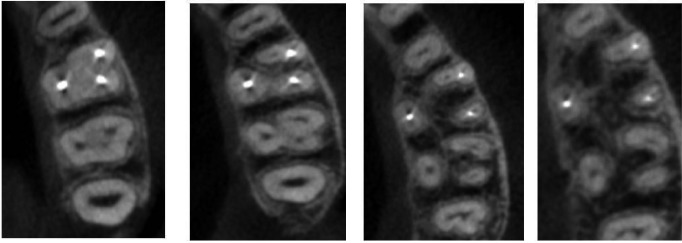
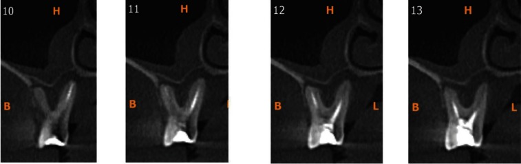
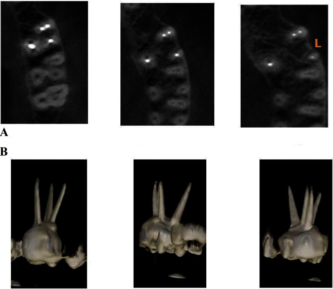
Material and methods: A retrospective study of 100 CBCTs of patients were underwent scanning for various treatment modalities, with asymptomatic endodontically treated permanent first and second maxillary molars were selected. History of root canal treatment varied from minimum of 1 year to a maximum of 10 years. Axial and paraxial images obtained were used to assess the presence of MB2 canal. Paraxial images were used to assess the periapical status.
Results: Of the 100 scans, 66 were of permanent maxillary first molar and 34 were of permanent maxillary second molar. The incidence of MB2 canal was 86.36% in maxillary first molars and 29.4% in maxillary second molars. 77.19 % of maxillary first molars and 90% of maxillary second molars had an unfilled MB2 canal. 72.7% of maxillary first molars and 88.8% of maxillary second molars showed significant periapical radiolucencies in unfilled MB2 canals.
Conclusions: MB2 canals were present in majority of cases and most of the unfilled MB2 canals showed evidence of periapical radiolucencies. Key words:MB2 Canals, Cone Beam computed Tomography (CBCT), Filled /Unfilled canals, Endodontically treated teeth.
Conflict of interest statement
None.
Figures
References
-
- Peters OA. Current challenges and concepts in the preparation of root canal systems: a review. J Endod. 2004;30:559–67. - PubMed
-
- Pattanshetti N, Gaidhene M, Al Kandari AM. Root canal morphology of the mesiobuccal and distal roots of permanent first molars in a Kuwait population. A clinical study. Int Endod J. 2008;41:755–62. - PubMed
-
- Weine FS, Healey HJ, Gerstein H, Evanson L. Canal configuration in the mesiobuccal root of the maxillary first molar and its endodontic significance. Oral Surg Oral Med Oral Pathol. 1969;28:419–25. - PubMed
-
- Weller RN, Hartwell GR. The impact of improved access and searching techniques on detection of the mesiolingual canal in maxillary molars. J Endod. 1989;15:82–3. - PubMed
-
- Wasti F, Shearer AC, Wilson NH. Root canal systems of the mandibular and maxillary first permanent molar teeth of south Asian Pakistanis. Int Endod J. 2001;34:263–6. - PubMed
LinkOut - more resources
Full Text Sources
Other Literature Sources