Review
. 2016 Dec;8(12):3762-3773.
doi: 10.21037/jtd.2016.12.88.

Veno-venous extracorporeal membrane oxygenation: cannulation techniques

Affiliations
Review

Veno-venous extracorporeal membrane oxygenation: cannulation techniques

Carlo Banfi et al. J Thorac Dis. 2016 Dec.

Abstract

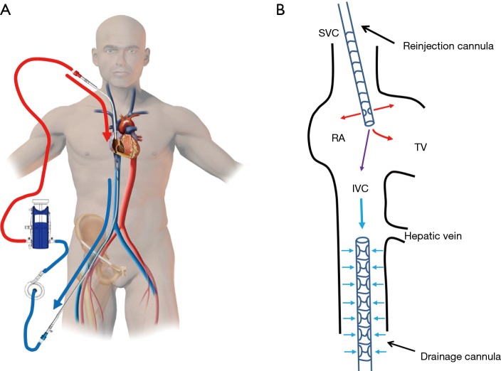
The development of extracorporeal membrane oxygenation (ECMO) technology allows a new approach for the intensive care management of acute cardiac and/or respiratory failure in adult patients who are not responsive to conventional treatment. Current ECMO therapies provide a variety of options for the multidisciplinary teams who are involved in the management of these critically ill patients. In this regard, veno-venous ECMO (VV-ECMO) can provide quite complete respiratory support, even if this highly complex technique presents substantial risks, such as bleeding, thromboembolic events and infection. While VV-ECMO circuits usually include the cannulation of two vessels (double cannulation) in its classic configuration, the use of a single cannula is now possible for VV-ECMO support. Recently, experienced centers have employed more advanced approaches by cannulating three vessels (triple cannulation) which follows veno-arterio-venous (VAV) or veno-arterio-pulmonary-arterial cannulation (VAPa). However, 'triple' cannulation expands the field of application but increases the complexity of ECMO systems. In the present review, the authors focus on the indications for VV-ECMO, patient assessment prior to cannulation, the role of ultrasound-guided vessel puncture, double lumen single bicaval cannulations, and finally triple cannulation in VV-ECMO.

Keywords: Respiratory failure; veno-venous extracorporeal membrane oxygenation (VV-ECMO).


Conflict of interest statement

The authors have no conflicts of interest to declare.

Figures

Figure 1
Figure 1
Classical double VV-ECMO cannulation. (A) Femoro-jugular VV-ECMO; (B) the venous drainage is achieved via a large multiport cannula introduced into the femoral vein and advanced at the junction between the inferior vena cava (IVC) and the right atrium (RA); the reinjection is performed via an arterial cannula introduced into the right internal jugular vein and advanced through the superior vena cava (SVC) into the RA, in front of the tricuspid valve (TV). VV-ECMO, veno-venous extracorporeal membrane oxygenation.
Figure 2
Figure 2
Transesophageal echocardiography (mid-esophageal 0° four-chamber view). The tip of the reinjection cannula is located in the right atrium (RA) in front of the tricuspid valve (TV). RV, right ventricle.
Figure 3
Figure 3
Transesophageal echocardiography (transgastric 65° view). The tip of the drainage cannula is located in the inferior vena cava (IVC) at the junction between the IVC and the hepatic vein.
Figure 4
Figure 4
Chest X-ray showing the correct distance between drainage and reinjection cannulae.
Figure 5
Figure 5
A: the 180° manufactured curved tube diminish the shear stress and the risk of kinking on the reinjection cannula; B: the fixation of the return cannula on the mastoid.
Figure 6
Figure 6
Double VV-ECMO cannulation. (A) Femoro-femoral VV-ECMO; (B) the venous drainage is accomplished via a large multiport cannula introduced into the femoral vein and advanced to the mid-IVC; the reinjection uses a cannula introduced into the contralateral femoral vein and advanced to the RA. VV-ECMO, veno-venous extracorporeal membrane oxygenation; TV, tricuspid valve; IVC, inferior vena cava; RA, right atrium; SVC, superior vena cava.
Figure 7
Figure 7
Dual-lumen single bicaval cannulation. (A) Avalon (Maquet, Rastatt, Germany) dual-lumen single bicaval cannulation technique; (B) drainage and reinjection are assured via a double-lumen cannula introduced into the right internal jugular vein. The cannula is advanced until the tip lies in the mid-IVC, in front of the hepatic veins. Drainage is from the IVC and the SVC. Reinjection is into the RA facing to the tricuspid valve. VV-ECMO, veno-venous extracorporeal membrane oxygenation; TV, tricuspid valve; IVC, inferior vena cava; RA, right atrium; SVC, superior vena cava.
Figure 8
Figure 8
Veno-arterio-venous extracorporeal membrane oxygenation (VAV-ECMO).

References

    1. Abrams D, Brodie D. Novel Uses of Extracorporeal Membrane Oxygenation in Adults. Clin Chest Med 2015;36:373-84. 10.1016/j.ccm.2015.05.014 - DOI - PubMed
    1. Combes A, Brodie D, Bartlett R, et al. Position paper for the organization of extracorporeal membrane oxygenation programs for acute respiratory failure in adult patients. Am J Respir Crit Care Med 2014;190:488-96. 10.1164/rccm.201404-0630CP - DOI - PubMed
    1. Cheng R, Hachamovitch R, Kittleson M, et al. Complications of extracorporeal membrane oxygenation for treatment of cardiogenic shock and cardiac arrest: a meta-analysis of 1,866 adult patients. Ann Thorac Surg 2014;97:610-6. 10.1016/j.athoracsur.2013.09.008 - DOI - PubMed
    1. Ius F, Sommer W, Tudorache I, et al. Veno-veno-arterial extracorporeal membrane oxygenation for respiratory failure with severe haemodynamic impairment: technique and early outcomes. Interact Cardiovasc Thorac Surg 2015;20:761-7. 10.1093/icvts/ivv035 - DOI - PubMed
    1. Bartlett RH, Deatrick KB. Current and future status of extracorporeal life support for respiratory failure in adults. Curr Opin Crit Care 2016;22:80-5. 10.1097/MCC.0000000000000274 - DOI - PubMed

LinkOut - more resources