

Repair of bone defects with prefabricated vascularized bone grafts and double-labeled bone marrow-derived mesenchymal stem cells in a rat model
- PMID: 28150691
- PMCID: PMC5288698
- DOI: 10.1038/srep39431
Repair of bone defects with prefabricated vascularized bone grafts and double-labeled bone marrow-derived mesenchymal stem cells in a rat model
Erratum in
-
Author Correction: Repair of bone defects with prefabricated vascularized bone grafts and double-labeled bone marrow-derived mesenchymal stem cells in a rat model.Sci Rep. 2020 Jul 30;10(1):12863. doi: 10.1038/s41598-020-69955-3. Sci Rep. 2020. PMID: 32732986 Free PMC article.
Abstract
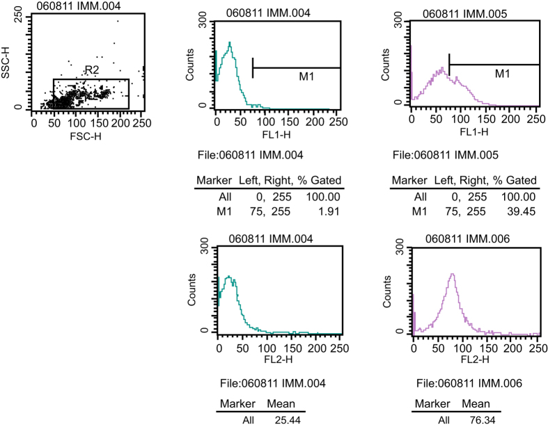
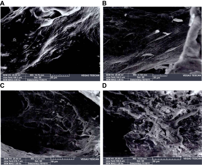
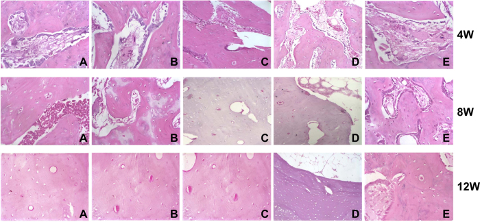
This study aims to investigate the repair of bone defects with prefabricated vascularized bone grafts and double-labeled bone marrow-derived mesenchymal stem cells (BMSCs) in a rat model. BMSCs were separated from rat bone marrow. LTR-CMVpro-RFP and LTR-CMVpro-GFP were transfected into the BMSCs for in vitro and in vivo tracking. BMSCs-RFP and BMSCs-GFP were induced into endothelial progenitor cells (EPCs) and osteoblasts (OBs). Rats were divided into five groups: Group A: in vitro prefabrication with EPCs-RFP + in vivo prefabrication with arteriovenous vascular bundle + secondary OBs-GFP implantation; Group B: in vitro prefabrication with EPCs-RFP + secondary OBs-GFP implantation; Group C: in vivo prefabrication with arteriovenous vascular bundle + secondary OBs-GFP implantation; Group D: implantation of EPCs-RFP + implantation of with arteriovenous vascular bundle + simultaneous OBs-GFP implantation; Group E: demineralized bone matrix (DBM) grafts (blank control). Among five groups, Group A had the fastest bone regeneration and repair, and the regenerated bone highly resembled normal bone tissues; Group D also had fast bone repair, but the repair was slightly slower than Group A. Therefore, in vitro prefabrication with EPCs-RFP plus in vivo prefabrication with arteriovenous vascular bundle and secondary OBs-GFP implantation could be the best treatment for bone defect.
Conflict of interest statement
The authors declare no competing financial interests.
Figures
Similar articles
-
Adenovirus-Mediated Expression of BMP-2 and BFGF in Bone Marrow Mesenchymal Stem Cells Combined with Demineralized Bone Matrix For Repair of Femoral Head Osteonecrosis in Beagle Dogs.Cell Physiol Biochem. 2017;43(4):1648-1662. doi: 10.1159/000484026. Epub 2017 Oct 18. Cell Physiol Biochem. 2017. PMID: 29045937
-
Endothelial progenitor cells improve the therapeutic effect of mesenchymal stem cell sheets on irradiated bone defect repair in a rat model.J Transl Med. 2018 May 22;16(1):137. doi: 10.1186/s12967-018-1517-4. J Transl Med. 2018. PMID: 29788957 Free PMC article.
-
Prefabrication of a vascularized bone graft with Beta tricalcium phosphate using an in vivo bioreactor.Artif Organs. 2013 Oct;37(10):884-93. doi: 10.1111/aor.12092. Epub 2013 May 6. Artif Organs. 2013. PMID: 23646847
-
Stem Cells for Bone Regeneration: Current State and Future Directions.J Craniofac Surg. 2019 May/Jun;30(3):730-735. doi: 10.1097/SCS.0000000000005250. J Craniofac Surg. 2019. PMID: 30817525 Review.
-
Bone Graft Prefabrication Following the In Vivo Bioreactor Principle.EBioMedicine. 2016 Oct;12:43-54. doi: 10.1016/j.ebiom.2016.09.016. Epub 2016 Sep 20. EBioMedicine. 2016. PMID: 27693103 Free PMC article. Review.
Cited by
-
Ectopic transient overexpression of OCT-4 facilitates BMP4-induced osteogenic transdifferentiation of human umbilical vein endothelial cells.J Tissue Eng. 2020 Mar 7;11:2041731420909208. doi: 10.1177/2041731420909208. eCollection 2020 Jan-Dec. J Tissue Eng. 2020. PMID: 32201555 Free PMC article.
-
Material-Induced Venosome-Supported Bone Tubes.Adv Sci (Weinh). 2019 Jul 1;6(17):1900844. doi: 10.1002/advs.201900844. eCollection 2019 Sep 4. Adv Sci (Weinh). 2019. PMID: 31508287 Free PMC article.
-
Insulin growth factor-1 promotes the proliferation and osteogenic differentiation of bone marrow mesenchymal stem cells through the Wnt/β-catenin pathway.Exp Ther Med. 2021 Aug;22(2):891. doi: 10.3892/etm.2021.10323. Epub 2021 Jun 17. Exp Ther Med. 2021. PMID: 34194569 Free PMC article.
-
Restoration of mandibular bone defects with demineralized bone matrix combined with three-dimensional cultured bone marrow-derived mesenchymal stem cells in minipig models.J Mater Sci Mater Med. 2018 Aug 31;29(9):147. doi: 10.1007/s10856-018-6152-3. J Mater Sci Mater Med. 2018. PMID: 30171486
-
Cellular direct conversion by cell penetrable OCT4-30Kc19 protein and BMP4 growth factor.Biomater Res. 2022 Jul 14;26(1):33. doi: 10.1186/s40824-022-00280-8. Biomater Res. 2022. PMID: 35836274 Free PMC article.
References
-
- Christensen B. B. Autologous tissue transplantations for osteochondral repair. Dan Med J 63 (2016). - PubMed
-
- Askari A., Farjam M. & Zohalinezhad M. E. Early reports of bone repair techniques and bone xenograft in Persian traditional medicine. J Integr Med 13, 140–141 (2015). - PubMed
-
- Ren M. L. et al.. Allogeneic adipose-derived stem cells with low immunogenicity constructing tissue-engineered bone for repairing bone defects in pigs. Cell Transplant 21, 2711–2721 (2012). - PubMed
Publication types
MeSH terms
Substances
LinkOut - more resources
Full Text Sources
Other Literature Sources
Medical
Molecular Biology Databases
