. 2017 Feb 2:7:41607.
doi: 10.1038/srep41607.

Bioengineering a bacterial pathogen to assemble its own particulate vaccine capable of inducing cellular immunity

Affiliations

Bioengineering a bacterial pathogen to assemble its own particulate vaccine capable of inducing cellular immunity

Jason W Lee et al. Sci Rep. .

Abstract

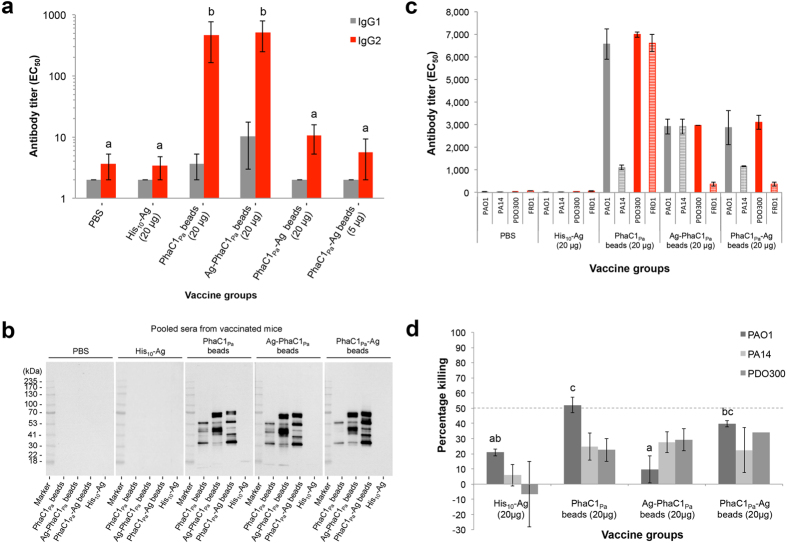
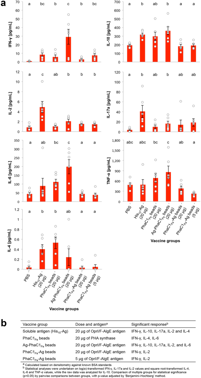
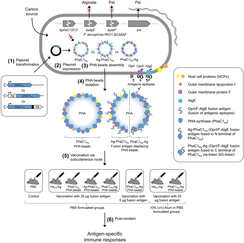
Many bacterial pathogens naturally form cellular inclusions. Here the immunogenicity of polyhydroxyalkanoate (PHA) inclusions and their use as particulate vaccines delivering a range of host derived antigens was assessed. Our study showed that PHA inclusions of pathogenic Pseudomonas aeruginosa are immunogenic mediating a specific cell-mediated immune response. Protein engineering of the PHA inclusion forming enzyme by translational fusion of epitopes from vaccine candidates outer membrane proteins OprI, OprF, and AlgE mediated self-assembly of PHA inclusions coated by these selected antigens. Mice vaccinated with isolated PHA inclusions produced a Th1 type immune response characterized by antigen-specific production of IFN-γ and IgG2c isotype antibodies. This cell-mediated immune response was found to be associated with the production of functional antibodies reacting with cells of various P. aeruginosa strains as well as facilitating opsonophagocytic killing. This study showed that cellular inclusions of pathogenic bacteria are immunogenic and can be engineered to display selected antigens suitable to serve as particulate subunit vaccines against infectious diseases.


Conflict of interest statement

B.H.A.R. is founding inventor and shareholder of PolyBatics Ltd.

Figures

Figure 1
Figure 1. A schematic of the generation of P. aeruginosa knockout mutant PAO1 ΔCΔ8ΔF.
In order to promote the production of PHAMCL inclusions and vaccine candidate EPS Psl, site-directed homologous recombination was used to delete major parts of (a) alg8 and (b) pelF genes encoding a glycosyltransferase in the PHA negative mutant PAO1 ΔphaC1ZC2. (c) Resultant triple mutant strain is defective in PHA/alginate/pel polysaccharide was verified by DNA sequencing (see Supplementary Fig. 1).
Figure 2
Figure 2. Assessment of the tolerance of the class II PHA synthase (PhaC1Pa) to C terminal fusion.
(a) Schematic representation of fusion protein PhaC1Pa-GFP for assessment of class II PHA synthase tolerance to C terminal fusion. (b) Fluorescence microscopy analysis of Nile red stained P. aeruginosa PAO1 ΔCΔ8ΔF cultures harboring various plasmids grown under PHAMCL accumulating conditions for 24 h and visualized for GFP and in vivo PHAMCL inclusions (white arrow). (c) Quantification and compositional analysis of PHAMCL in whole-cell by Gas chromatography-mass spectrometry (GC/MS). (d) SDS-PAGE and immunoblot analysis of cell lysates to confirm the production of fusion protein. ΔCΔ8ΔF + MCS-5, PAO1 ΔCΔ8ΔF + pBBR1MCS-5; ΔCΔ8ΔF + JO-5_C1, PAO1 ΔCΔ8ΔF + pBBR1JO-5_C1; ΔCΔ8ΔF + JO-5_C1gfp, PAO1 ΔCΔ8ΔF + pBBR1JO-5_C1gfp; ND, not detected; 3-HH (C6), methyl 3-hydroxyhexanoate; 3-HO (C8), methyl 3-hydroxyoctanoate; 3-HN (C9), methyl 3-hydroxynonanoate; 3-HD (C10), methyl 3-hydroxydecanoate; 3-HUD (C11), methyl 3-hydroxyundecanoate; 3-HDD isomer (C12), methyl 3-hydroxydodecanoate isomer; 3-HDD (C12), methyl 3-hydroxydodecanoate; 3-HTD (C14), methyl 3-hydroxytetradecanoate.
Figure 3
Figure 3. Bioengineering and production of vaccine PHAMCL inclusions in vivo.
PAO1 ΔCΔ8ΔF cells harboring various plasmids cultivated under PHAMCL accumulating condition for 48 h and their respective isolated PHAMCL beads. (a) A schematic representation of various fusion proteins which mediate PHAMCL bead production in strain PAO1 ΔCΔ8ΔF or recombinant protein production in E. coli (see Supplementary Fig. 3a,b). (b) Accumulation and size of PHAMCL inclusions were analysised by Transmission Electron Microscopy (TEM) in whole-cells and of the isolated PHAMCL bead material. (c) Quantification and compositional analysis of PHAMCL using GC/MS. BDW, percentage of the bead dry weight; ΔCΔ8ΔF + MCS-5, PAO1 ΔCΔ8ΔF + pBBR1MCS-5; ΔCΔ8ΔF + JO-5_C1, PAO1 ΔCΔ8ΔF + pBBR1JO-5_C1; ΔCΔ8ΔF + JO-5_C1gfp, PAO1 ΔCΔ8ΔF + pBBR1JO-5_C1gfp; ND, not detected; 3-HH (C4), methyl 3-hydroxybutanoate; 3-HH (C6), methyl 3-hydroxyhexanoate; 3-HO (C8), methyl 3-hydroxyoctanoate; 3-HN (C9), methyl 3-hydroxynonanoate; 3-HD (C10), methyl 3-hydroxydecanoate; 3-HUD (C11), methyl 3-hydroxyundecanoate; 3-HDD isomer (C12), methyl 3-hydroxydodecanoate isomer; 3-HDD (C12), methyl 3-hydroxydodecanoate; 3-HTD (C14), methyl 3-hydroxytetradecanoate; 3-HHD (C16), methyl 3-hydroxyhexadecanote.
Figure 4
Figure 4. Protein analysis of vaccine PHAMCL beads.
(a) SDS-PAGE analysis of whole-cell lysate and PHAMCL beads stained with Coomassie Blue or (b) probed using anti-PhaC1 polyclonal antibodies raised against various epitopes of PhaC1Pa (see Materials). Fusion proteins of interest in PHAMCL bead samples are indicated by black arrows and the eleven HCP bands identified of interest are indicated in roman numerals. These indicated proteins were isolated for protein identification by MALDI-TOF MS (see Supplementary Tables 1 and 2). Detected bands from b immunoblotting were overlaid and matched to specific bands on the Coomassie Blue stained gel (red square). Antibody detected epitopes were aligned with peptides identified by MALDI-TOF MS (see Supplementary Table 3). Major copurified protein bands from PHAMCL beads formed by PHA synthase antigen fusions were identified on SDS-PAGE (see Materials for criteria). (c) Immunoblot analysis of isolated PHAMCL beads using polyclonal antibodies raised against epitopes of OprF or OprI or AlgE. (d) Table summarizing the Identification of the eleven PHAMCL bead associated HCPs by MALDI-TOF MS (see also Supplementary Table 2). ΔCΔ8ΔF + MCS-5, PAO1 ΔCΔ8ΔF + pBBR1MCS-5; ΔCΔ8ΔF + JO-5_C1, PAO1 ΔCΔ8ΔF + pBBR1JO-5_C1; ΔCΔ8ΔF + JO-5_C1gfp, PAO1 ΔCΔ8ΔF + pBBR1JO-5_C1gfp.
Figure 5
Figure 5. Antibody response to vaccination with vaccine PHAMCL beads.
(a) Antigen-specific IgG1 or IgG2c isotype antibody responses measured by ELISA using a pool of OprI, OprF, and AlgE antigen specific peptides from sera. Results are expressed in reciprocal antibody titers, representing the dilution required to obtain half of the maximal amount of the OD signal (EC50). (b) To identify antigenic proteins on PHAMCL beads, sera obtained was pooled in to their respective groups and used as a primary antibody for detection of epitopes on PHAMCL beads separated by SDS-PAGE. (c) Reactivity of pooled immune sera to different P. aeruginosa strains using a whole-cell ELISA was used. Nonmucoid (grey bars) and mucoid (red bars) strains of P. aeruginosa were tested. (d) Opsonic killing of P. aeruginosa nonmucoid strains by serum from mice immunized with vaccine PHAMCL beads. Bar represents the mean percent killing of three replicates for PAO1 and PA14 or duplicates for PDO300 relative to sera of the PBS vaccinated control group, and error bars represents the s.e.m. Data of graph for IgG are reported as means ± s.e.m (6 mice per group). Statistical significance (p < 0.05) of IgG2c is indicated by ‘letter based’ representation of pairwise comparisons between groups using Tukey’s post-hoc test. IgG1 were not statistically significant. Data of graph for whole-cell ELISA represent means of two replicates of pooled sera ± s.e.m of the replicates. There are insufficient replicates to undertake a statistical analysis. Statistical significance (p < 0.05) for opsonic killing assay is indicated by ‘letter based’ representation of pairwise comparisons between groups using Tukey’s post-hoc test. PA14 were not statistically significant. There are insufficient replicates to undertake a statistical analysis for PDO300.
Figure 6
Figure 6. Cytokine response to vaccination with vaccine PHAMCL beads.
(a) Release of cytokines from splenocyte cultures restimulated with soluble recombinant His10-Ag was measured by cytometric bead array. Results are calculated by subtracting cytokine values of the media-stimulated samples from the cytokine values of the recombinant protein stimulated samples. (b) Summary of the cytokine response as a comparison to PBS negative control group. Data of graphs are reported as means ± s.e.m and each individual mouse are reported as a dot (n = 6 per group). Statistical significance (p < 0.05) is indicated by ‘letter-based’ representation of pairwise comparisons between groups.
Figure 7
Figure 7. Engineering the pathogens intrinsic ability to produce PHAMCL beads as particulate subunit vaccines.
A schematic overview of the production and immunological evaluation of custom-made PHAMCL beads displaying both engineered vaccine candidate antigens and antigens derived from the host expression cells. (1) Plasmid encoding wildtype PHA synthase (phaC1Pa) or OprI/F-AlgE fusion antigen fused to the N terminal of PhaC1Pa or OprI/F-AlgE fusion antigen fused to the C terminal of PhaC1Pa via linker-SG-linker were transformed in to P. aeruginosa PAO1 ΔCΔ8ΔF mutant strain. This strain is defective in production of native PHAMCL and of EPS alginate and Pel (see Fig. 1). (2) Plasmid harboring strains are then grown under PHAMCL accumulating conditions to mediate overproduction of the fusion protein and subsequent PHAMCL bead assembly (See Fig. 3a–c). (3) Formation of PHAMCL beads results in the display of fusion antigens covalently linked to the PHA synthase and the incorporation of granule associated and HCPs (See Fig. 4a–d). (4) PHAMCL beads are isolated from the host by mechanical disruption and subsequently purified. (5) C57BL/6 mice were vaccinated with sterilized PHAMCL beads, recombinant His-tagged OprI/F-AlgE, and PBS via subcutaneous route three times at biweekly intervals. (6) Blood and splenocytes were collected from mice euthanized three-weeks after the last vaccination for analysis. Antigen-specific serum antibodies (ELISA) (see Fig. 5a) and cytokines (Cytometric bead array, mouse Th1/Th2/Th17 cytokine kit) (see Fig. 6a,b) were measured.

Similar articles

Cited by

References

    1. Draper J. et al.. Polyhydroxyalkanoate inclusions: polymer synthesis, self-assembly and display technology. In: Bionanotechnology: biological self assembly and its applications (ed^(eds Rehm B. H. A.) Caister Academic Press (2013).
    1. Rehm B. H. A. Polyester synthases: natural catalysts for plastics. Biochemical Journal 376, 15–33 (2003). - PMC - PubMed
    1. Foged C. Subunit vaccines of the future: the need for safe, customized and optimized particulate delivery systems. Therapeutic delivery 2, 1057–1077 (2011). - PubMed
    1. Leleux J. & Roy K. Micro and nanoparticle‐based delivery systems for vaccine immunotherapy: an immunological and materials perspective. Advanced healthcare materials 2, 72–94 (2013). - PubMed
    1. Parlane N. A. et al.. Vaccines displaying mycobacterial proteins on biopolyester beads stimulate cellular immunity and induce protection against tuberculosis. Clinical and Vaccine Immunology 19, 37–44 (2012). - PMC - PubMed

Publication types

MeSH terms