Review
. 2017 Feb 1;10(2):89-104.
doi: 10.1242/dmm.027649.

Helicobacter pylori-induced gastric pathology: insights from in vivo and ex vivo models

Affiliations
Review

Helicobacter pylori-induced gastric pathology: insights from in vivo and ex vivo models

Michael D Burkitt et al. Dis Model Mech. .

Abstract

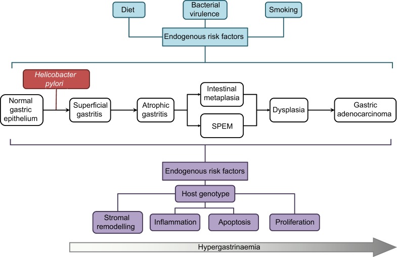
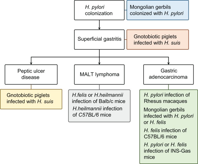
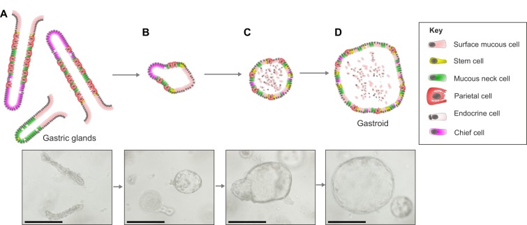
Gastric colonization with Helicobacter pylori induces diverse human pathological conditions, including superficial gastritis, peptic ulcer disease, mucosa-associated lymphoid tissue (MALT) lymphoma, and gastric adenocarcinoma and its precursors. The treatment of these conditions often relies on the eradication of H. pylori, an intervention that is increasingly difficult to achieve and that does not prevent disease progression in some contexts. There is, therefore, a pressing need to develop new experimental models of H. pylori-associated gastric pathology to support novel drug development in this field. Here, we review the current status of in vivo and ex vivo models of gastric H. pylori colonization, and of Helicobacter-induced gastric pathology, focusing on models of gastric pathology induced by H. pylori, Helicobacter felis and Helicobacter suis in rodents and large animals. We also discuss the more recent development of gastric organoid cultures from murine and human gastric tissue, as well as from human pluripotent stem cells, and the outcomes of H. pylori infection in these systems.

Keywords: Gastric cancer; Gastroid; Helicobacter; MALT lymphoma; Organoid; Peptic ulcer disease.


Conflict of interest statement

Competing interests

The authors declare no competing or financial interests.

Figures

Fig. 1.
Fig. 1.
Worldwide prevalence of Helicobacter pylori infection. The map shows the prevalence of H. pylori infection in different parts of the world. Note, the particularly high prevalence in sub-Saharan Africa, Latin America and the Middle East. Australasia, Switzerland, and more generally North America and Western Europe have the lowest incidence of H. pylori infection. Data derived from Asfeldt et al., 2008; Ben Mansour et al., 2016; Laszewicz et al., 2014; Luzza et al., 2014; McDonald et al., 2015; Peleteiro et al., 2014; Saltanova, 2001; Sanchez Ceballos et al., 2007; van Blankenstein et al., 2013.
Fig. 2.
Fig. 2.
The anatomy of the human and mouse stomach. A schematic of the anatomy of the human and mouse stomach and the structure of gastric glands. Two types of columnar mucosa line the human stomach: the antrum is lined with antral glands, whilst the corpus and fundus are lined with deeper oxyntic, or corpus glands (see Glossary, Box 1). The murine stomach has areas that are analogous to the human stomach, including antral and corpus glands, and it also has a forestomach lined with squamous epithelium. Stem cells that reside at the base of the gland generate the antral gland. Following asymmetric cell division in the stem cell zone, daughter cells migrate upwards towards the gastric lumen and differentiate into mucous neck, surface mucous and endocrine cells. In corpus glands, the stem cell niche is located at the isthmus of the gland. Cells migrate upwards from the stem cell zone and differentiate into surface mucous cells. Other cells migrate down the gland and differentiate into acid-secreting parietal cells, endocrine cells, or zymogen-secreting (see Glossary, Box 1) chief cells.
Fig. 3.
Fig. 3.
H. pylori infection and progression to gastric cancer. A schematic demonstrating the pathological progression of H. pylori-induced gastric pre-neoplasia, and highlighting endogenous risk factors for progression towards gastric cancer. SPEM, spasmolytic polypeptide-expressing metaplasia.
Fig. 4.
Fig. 4.
Modelling the pathological outcomes of Helicobacter infection. A schematic of the principal pathological outcomes of Helicobacter infection in humans, annotated with details of the best-characterized in vivo models for these conditions.
Fig. 5.
Fig. 5.
Gastric organoid culture and differentiation. Diagrams and images showing the maturation of gastric organoids. (A) Freshly digested gastric corpus glands from a C57BL/6 mouse. (B) Glands 24 h after harvesting that have formed immature organoids and have a small spherical appearance. (C) On day 3 of culture, the immature organoids have expanded and can be passaged. (D) Following passage, the organoids retain their spherical appearance and continue to grow. Images from authors' own laboratories. Scale bars: 250 µm.

References

    1. Agha A. and Graham D. Y. (2005). Evidence-based examination of the African enigma in relation to Helicobacter pylori infection. Scand. J. Gastroenterol. 40, 523-529. 10.1080/00365520510012280 - DOI - PubMed
    1. Amorim I., Taulescu M. A., Day M. J., Catoi C., Reis C. A., Carneiro F. and Gärtner F. (2016). Canine gastric pathology: a review. J. Comp. Pathol. 154, 9-37. 10.1016/j.jcpa.2015.10.181 - DOI - PubMed
    1. Andrews P. L., Illman O. and Mellersh A. (1979). Some observations of anatomical abnormalities and disease states in a population of 350 ferrets (Mustela furo L.). Z. Versuchstierkd. 21, 346-353. - PubMed
    1. Asenjo L. M. and Gisbert J. P. (2007). [Prevalence of Helicobacter pylori infection in gastric MALT lymphoma: a systematic review]. Rev. Esp. Enferm. Dig. 99, 398-404. 10.4321/S1130-01082007000700006 - DOI - PubMed
    1. Asfeldt A. M., Straume B., Steigen S. E., Løchen M.-L., Florholmen J., Bernersen B., Johnsen R. and Paulssen E. J. (2008). Changes in the prevalence of dyspepsia and Helicobacter pylori infection after 17 years: the Sørreisa gastrointestinal disorder study. Eur. J. Epidemiol. 23, 625-633. 10.1007/s10654-008-9275-x - DOI - PubMed