. 2017 Aug;19(8):900-908.
doi: 10.1038/gim.2016.211. Epub 2017 Feb 2.

Clinical and molecular consequences of disease-associated de novo mutations in SATB2

Affiliations

Clinical and molecular consequences of disease-associated de novo mutations in SATB2

Hemant Bengani et al. Genet Med. 2017 Aug.

Abstract

Purpose: To characterize features associated with de novo mutations affecting SATB2 function in individuals ascertained on the basis of intellectual disability.

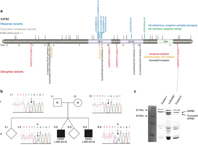
Methods: Twenty previously unreported individuals with 19 different SATB2 mutations (11 loss-of-function and 8 missense variants) were studied. Fibroblasts were used to measure mutant protein production. Subcellular localization and mobility of wild-type and mutant SATB2 were assessed using fluorescently tagged protein.

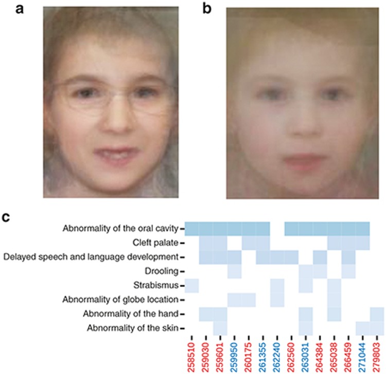
Results: Recurrent clinical features included neurodevelopmental impairment (19/19), absent/near absent speech (16/19), normal somatic growth (17/19), cleft palate (9/19), drooling (12/19), and dental anomalies (8/19). Six of eight missense variants clustered in the first CUT domain. Sibling recurrence due to gonadal mosaicism was seen in one family. A nonsense mutation in the last exon resulted in production of a truncated protein retaining all three DNA-binding domains. SATB2 nuclear mobility was mutation-dependent; p.Arg389Cys in CUT1 increased mobility and both p.Gly515Ser in CUT2 and p.Gln566Lys between CUT2 and HOX reduced mobility. The clinical features in individuals with missense variants were indistinguishable from those with loss of function.

Conclusion: SATB2 haploinsufficiency is a common cause of syndromic intellectual disability. When mutant SATB2 protein is produced, the protein appears functionally inactive with a disrupted pattern of chromatin or matrix association.Genet Med advance online publication 02 February 2017.


Figures

Figure 1
Figure 1
SATB2 mutation spectrum and consequence. (a) The relative positions of the de novo mutation identified in this article are indicated in a cartoon representation of the SATB2 wild-type protein, which includes the location of the DNA-binding domains. Below the protein diagram are the mutations that are predicted to result in disruption of the open reading frame. The mutations are color-coded by type: nonsense (red), frameshift (purple), and essential splice site (orange). The intron–exon boundaries of the gene encoding the protein are shown as vertical gray lines and the exon number given between these. Above are the missense variants, which have been color-coded to represent the predicted effect of the residue substitution. Immediately above the protein cartoon is a density plot representing the position of all missense variants seen more than once in the Exome Aggregation Consortium database. (b) The family structure and cognate chromatographs are shown of the family with apparent gonadal mosaicism. The affected boys (II:2 29089 and II:4 29090) share the same essential splice site mutation, which is absent in both parents (I:1 and I:2). (c) Photograph of a western blot of protein derived from cultured fibroblasts of two unrelated control individuals and case 14 with a nonsense mutation in the final exon using an antibody made for the C-terminal region of SATB2. A pre-stained protein ladder (Invitrogen LC5925) was used to assess the size of the bands. The same membrane was imaged using both white light and chemiluminescence and the cropped ladder image was size matched and aligned to the immunoblot. The expected SATB2 band of ~80 kDa is seen in all three, with a band of unknown identity above this. A lower band, consistent with the production of the 692-amino-acid C-terminally truncated version on SATB2, is seen in case 14 but not in the control.
Figure 2
Figure 2
Effect of mutation within DNA-binding domains of SATB2. In silico predictions of the structural effects of missense mutations affecting CUT1 (a) or CUT2 (b) are shown. In each case, the boundaries of alpha helixes 3 and 2 have been altered (red arrows). The position of the cognate mutation within the DNA-binding domain is shown on a cartoon of the SATB2 protein in d. (c) Western blot analysis of fractionated HEK293 cells with Tetracyclin (TET) inducible expression of GFP-tagged mutant and wild-type SATB2. Most SATB2 is attached to the matrix. The CUT1 missense mutation results in a marked increase in the proportion of the protein in the soluble fraction. The missense mutation between CUT2 and HOX shows apparent reduced levels of matrix association. Antibodies against lamin and histone are presented to show that the fractionation has worked. (d) The gray-shaded box represents the protein product of an artificial control mutation that has been made in the cDNA of SATB2 to create a peptide with none of the DNA-binding domains. This is termed the “N-terminal region.” (e) Confocal photomicrographs of cells transiently transfected with plasmids containing wild-type and mutant GFP-tagged SATB2 cDNA under the control of a ubiquitous promoter. The mutation is shown above each photograph. The wild-type, p.Gly515Ser, and p.Gln566Lys cDNAs all produce a granular pattern of fluorescence within the nucleus, the N-terminal regions show a punctate pattern, and the p.Arg389Cys shows a glassy diffuse pattern. (f, g) Representative images of cells from independent transient transfections of the wild-type, p.Arg389Cys, p.Gly515Ser, and p.Gln566Lys cDNAs to assess fluorescence loss and recovery after multiple photobleaching using regions of the nucleus (illustrated for this experiment by the red squares on the photomicrographs). This graph shows quantitation of the fluorescence after each photobleach and recovery. The error bars represent standard deviations for the measurements at each time point.
Figure 3
Figure 3
Facial characteristics associated with de novo mutations in SATB2. (a) Average face constructed from the available published facial images from individuals with de novo mutations in SATB2. (b) Average face constructed from facial photographs from 10 of the individuals with the de novo mutation in SATB2 reported here. Both images show a small mouth with a thin upper lip and facial asymmetry. The asymmetry is subtle and most noticeable in the jaw line and corners of the mouth. (c) Heat map of the recurrently reported human phenotype ontology terms in the 14 Deciphering Developmental Disorders study individuals reported here in whom these terms had been systematically collected prior to the molecular diagnosis being made.
Figure 4
Figure 4
Provisional model for the role of SATB2 DNA-binding domains. Cartoon representation of SATB2 interacting with chromatin in vivo. This model shows the binding of CUT1 as an initiating and stabilizing event and CUT2 binding with an antagonistic or destabilizing event that promotes mobility of SATB2 within the nucleus. Uncertainty remains regarding the role of the HOX domain and whether the chromatin that binds to CUT1 and CUT2 is in cis or in trans with each other. The regions of the protein that mediate matrix attachment are not well defined, but a variant between the CUT2 and HOX domains appears to result in a reduced proportion of the SATB2 being matrix-attached.

References

    1. Brewer CM, Leek JP, Green AJ, et al. A locus for isolated cleft palate, located on human chromosome 2q32. Am J Hum Genet 1999;65:387–396. - PMC - PubMed
    1. FitzPatrick DR, Carr IM, McLaren L, et al. Identification of SATB2 as the cleft palate gene on 2q32-q33. Hum Mol Genet 2003;12:2491–2501. - PubMed
    1. Large-scale discovery of novel genetic causes of developmental disorders. Nature. 2015;519:223–228. - PMC - PubMed
    1. Dobreva G, Chahrour M, Dautzenberg M, et al. SATB2 is a multifunctional determinant of craniofacial patterning and osteoblast differentiation. Cell 2006;125:971–986. - PubMed
    1. Alcamo EA, Chirivella L, Dautzenberg M, et al. Satb2 regulates callosal projection neuron identity in the developing cerebral cortex. Neuron 2008;57:364–377. - PubMed

Publication types

MeSH terms

Substances