. 2017 Feb 2;12(2):e0171215.
doi: 10.1371/journal.pone.0171215. eCollection 2017.

Highly tumorigenic hepatocellular carcinoma cell line with cancer stem cell-like properties

Affiliations

Highly tumorigenic hepatocellular carcinoma cell line with cancer stem cell-like properties

Benoit Lacoste et al. PLoS One. .

Abstract

There are limited numbers of models to study hepatocellular carcinoma (HCC) in vivo in immunocompetent hosts. In an effort to develop a cell line with improved tumorigenicity, we derived a new cell line from Hepa1-6 cells through an in vivo passage in C57BL/6 mice. The resulting Dt81Hepa1-6 cell line showed enhanced tumorigenicity compared to Hepa1-6 with more frequent (28±12 vs. 0±0 lesions at 21 days) and more rapid tumor development (21 (100%) vs. 70 days (10%)) in C57BL/6 mice. The minimal Dt81Hepa1-6 cell number required to obtain visible tumors was 100,000 cells. The Dt81Hepa1-6 cell line showed high hepatotropism with subcutaneous injection leading to liver tumors without development of tumors in lungs or spleen. In vitro, Dt81Hepa1-6 cells showed increased anchorage-independent growth (34.7±6.8 vs. 12.3±3.3 colonies; P<0.05) and increased EpCAM (8.7±1.1 folds; P<0.01) and β-catenin (5.4±1.0 folds; P<0.01) expression. A significant proportion of Dt81Hepa1-6 cells expressed EpCAM compared to Hepa1-6 (34.8±1.1% vs 0.9±0.13%; P<0.001). Enriched EpCAM+ Dt81Hepa1-6 cells led to higher tumor load than EpCAM- Dt81Hepa1-6 cells (1093±74 vs 473±100 tumors; P<0.01). The in vivo selected Dt81Hepa1-6 cell line shows high liver specificity and increased tumorigenicity compared to Hepa1-6 cells. These properties are associated with increased expression of EpCAM and β-catenin confirming that EpCAM+ HCC cells comprise a subset with characteristics of tumor-initiating cells with stem/progenitor cell features. The Dt81Hepa1-6 cell line with its cancer stem cell-like properties will be a useful tool for the study of hepatocellular carcinoma in vivo.


Conflict of interest statement

Competing Interests: The Novartis/Canadian Liver Foundation Hepatology Research Chair at the Université de Montréal is a philanthropic chair administered by Université de Montréal that was initially founded through a joint initiative of the Canadian Liver Foundation and Novartis to help promote research in the field of liver disease. There are no patents, products in development or marketed products to declare. This does not alter our adherence to PLOS ONE policies on sharing data and materials.

Figures
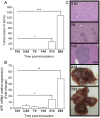
Fig 1
Fig 1. Dt81Hepa1-6 cell transfer leads to rapid tumor development.
A suspension of 1M Dt81Hepa1-6 cells was injected intrasplenically in C57BL/6 mice and animals were sacrificed after 3.5 to 28 days. (A) Manual count of tumor >0.5mm. (B) Alpha-fetoprotein (AFP) (Afp) mRNA expression in liver homogenates as measured by qPCR. (C) Representative microphotographs at 100x magnification of HPS-stained liver slices obtained after 3.5, 7 and 14 days. Whole-liver photographs obtained after 21 or 28 days. Control mice were injected with 1M Hepa1-6 cells and sacrificed at each time point (3.5 to 28 days) and pooled for analysis (Ctrl). (*P<0.05; **P<0.01; ***P<0.001).
Fig 2
Fig 2. Dose-dependent tumor development following transfer of Dt81Hepa1-6 cells.
Suspensions of Dt81Hepa1-6 cells [1K to 1M] were injected intrasplenically in C57BL/6 mice and animals were sacrificed after 28 days. (A) Manual count of tumor >0.5mm. (B) AFP (Afp) mRNA expression in liver homogenates by qPCR. (C) Representative microphotographs at 100x magnification of HPS-stained liver slice obtained after injection with 1K and 10K cells. Whole liver photographs of mice injected with 0.1M, 0.5M or 1M cells. Control mice were injected with 1M Hepa1-6 cells. (*P<0.05; **P<0.01; ***P<0.001).
Fig 3
Fig 3. Dt81Hepa1-6 cells are specific to the liver.
A suspension of 1M Dt81Hepa1-6 cells was injected subcutaneously (SC) in the left flank of C57BL/6 mice and animals were sacrificed after 28 days. (A) AFP mRNA relative expression in control and SC injected C57BL/6 mice. (B) Whole liver photographs at time of sacrifice. HPS-stained slices representative microphotographs of (C) lungs and spleen after SC injection and (D) Lungs and spleen after IS injection. Representative microphotographs were obtained at 100x magnification. (*P<0.05).
Fig 4
Fig 4. Dt81Hepa1-6 in vitro neoplastic profile.
A cell aggregation assay was performed and (A) the number of cells per aggregate was calculated 24h after seeding both cell lines in non-adherent agar-coated wells. Representative microphotographs are shown. In order to evaluate anchorage-independent growth, a soft-agar colony formation assay was performed; (B) cell lines were seeded at 10K-cell concentration in soft-agar gel [0.3%] in order to form visible colonies and were counted 5 weeks later. Representative microphotographs of cells after 5 weeks are shown. The ability of independent cells to form colonies without cell-to-cell contact was assessed by a proliferative clonal colony assay; (C) cell lines were seeded at 3K/mL concentration to obtain isolated cells and left to grow as colonies over a 4 weeks period and counted. The potential for invasiveness was evaluated with a double-layer droplet cell invasion assay; (D) Cell lines [20K cells] were embedded in COL1 [3mg/mL] to form a double layered droplet and cells penetrating the outer COL1 layer were counted every 12h for 8 days. Area under the curves (AUC) of the time-dependent invasion was calculated and used for statistical analysis. Representative microphotographs are shown. For the wound-healing assays (WHA); (E) normal and COL1-embedded WHA were performed on confluent cells for 24h and length traveled was evaluated using ImageJ. For the cell-spreading assay, (F) Cell lines were seeded for 6h on plastic and COL1 and cells showing an elongated morphology were counted manually. (G) Cell doubling time for Dt81Hepa1-6 and parental Hepa1-6 cells. (H) Viability of Dt81Hepa1-6 and Hepa1-6 cells following in vitro exposure to cisplatin [25ug/mL] for 24h. (*P<0.05; **P<0.01; ***P<0.001).
Fig 5
Fig 5. EpCAM+ Dt81Hepa1-6 cells are highly tumorigenic.
Dt81Hepa1-6 cells were incubated with APC-tagged anti-CD326 (EpCAM) antibody and sorted by FACS as EpCAM+high and EpCAM+low Dt81Hepa1-6 cells. Hepa1-6, Dt81Hepa1-6, EpCAM+high and EpCAM+low were analyzed by (A) qPCR for EpCAM mRNA expression and (B) flow cytometry after labeling with APC-tagged anti-CD326 (EpCAM). Dt81Hepa1-6, EpCAM+low and EpCAM+high were injected intrasplenically in C57BL/6 mice and sacrificed after 21 days. (C) Tumor load (>0.5mm) was assessed and (D) AFP mRNA expression was compared to healthy liver controls. (*P<0.05; **P<0.01; ***P<0.001).
Fig 6
Fig 6. Expression of β-Catenin, Cyclin D1 and Integrins by Dt81Hepa1-6 cells.
All experiments were performed on freshly trypsinized cells prior to their injection in vivo. Genes or proteins of interest were analyzed in Hepa1-6, Dt81Hepa1-6, EpCAM+low and EpCAM+high cell lines by qRT-PCR and/or western blot. (A) β-Catenin (Ctnnb1) gene and (B) protein expression shown on immunoblot (C) Cyclin D1 (Ccnd1), (D) Integrin-alpha1 (Itga1) and (E) Integrin-β1 (Itgb1) mRNA expression. (*P<0.05; **P<0.01).

Similar articles

Cited by

References

    1. El-Serag HB, Rudolph KL. Hepatocellular carcinoma: epidemiology and molecular carcinogenesis. Gastroenterology. 2007;132(7):2557–76. 10.1053/j.gastro.2007.04.061 - DOI - PubMed
    1. Blonski W, Kotlyar DS, Forde KA. Non-viral causes of hepatocellular carcinoma. World J Gastroenterol. 2010;16(29):3603–15. Epub 2010/08/03. 10.3748/wjg.v16.i29.3603 - DOI - PMC - PubMed
    1. Whittaker S, Marais R, Zhu AX. The role of signaling pathways in the development and treatment of hepatocellular carcinoma. Oncogene. 2010;29(36):4989–5005. Epub 2010/07/20. 10.1038/onc.2010.236 - DOI - PubMed
    1. Heindryckx F, Colle I, Van Vlierberghe H. Experimental mouse models for hepatocellular carcinoma research. Int J Exp Pathol. 2009;90(4):367–86. Epub 2009/08/08. 10.1111/j.1365-2613.2009.00656.x - DOI - PMC - PubMed
    1. Schnater JM, Bruder E, Bertschin S, Woodtli T, de Theije C, Pietsch T, et al. Subcutaneous and intrahepatic growth of human hepatoblastoma in immunodeficient mice. J Hepatol. 2006;45(3):377–86. Epub 2006/06/20. 10.1016/j.jhep.2006.03.018 - DOI - PubMed