

Retinal vessel density from optical coherence tomography angiography to differentiate early glaucoma, pre-perimetric glaucoma and normal eyes
- PMID: 28152070
- PMCID: PMC5289421
- DOI: 10.1371/journal.pone.0170476
Retinal vessel density from optical coherence tomography angiography to differentiate early glaucoma, pre-perimetric glaucoma and normal eyes
Abstract
Purpose: To evaluate optic nerve vascular density using swept source optical coherence tomography angiography (OCTA) in patients with early primary open angle glaucoma (POAG), pre-perimetric glaucoma and normal eyes.
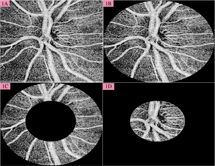
Methods: This is a prospective, observational study including 56 eyes in total and divided into 3 groups; 20 eyes with mild POAG, 20 pre-perimetric glaucoma eyes, and 16 age-matched normal eyes as controls. The optic disc region was imaged by a 1050-nm-wavelength swept-source OCT system (DRI OCT Triton, TOPCON). Vessel density was assessed as the ratio of the area occupied by the vessels in 3 distinct regions: 1) within the optic nerve head; 2) in the 3 mm papillary region around the optic disc; and 3) in the peripapillary region, defined as a 700-μm-wide elliptical annulus around the disc. The potential associations between vessel density and structural, functional measures were analyzed.
Results: There was a statistically significant difference for the peripapillary vessel density, optic nerve head vessel density, and papillary vessel density among all the groups (p<0.001). Control eyes showed a significant difference for all measured vessel densities compared to glaucomatous eyes (p values from 0.001 to 0.024). There was a statistically significant difference between control and pre-perimetric glaucoma eyes for peripapillary, optic nerve head and papillary vessel density values (p values from 0.001 to 0.007). The optic nerve head vessel density, superior and inferior papillary area vessel density (Pearson r = 0.512, 0.436, 0.523 respectively) were highly correlated with mean overall, superior and inferior RNFL thickness in POAG eyes (p = 0.04, p = 0.02 and p = 0.04 respectively). Multiple linear regression analysis of POAG group showed that optic nerve head vessel density in POAG group was more strongly linked to RNFL thickness than to any other variables.
Conclusions: Eyes with mild POAG could be differentiated from pre-perimetric glaucoma eyes, which also could be differentiated from normal eyes using OCTA-derived retinal vessel density measurements.
Conflict of interest statement
The authors have declared that no competing interests exist.
Figures
References
-
- Cantor LB, Wu Dunn D. Normal-tension glaucoma. In: Color Atlas of Glaucoma. 1998:155.
-
- Bonomi L, Marchini G, Marraffa M, Bernardi P, Morbio R, Varotto A. Vascular risk factors for primary open angle glaucoma: the Egna-Neumarkt Study. Ophthalmology 2000; 107:1287–93. - PubMed
Publication types
MeSH terms
Grants and funding
LinkOut - more resources
Full Text Sources
Other Literature Sources
