. 2017 Feb 2;13(2):e1006187.
doi: 10.1371/journal.ppat.1006187. eCollection 2017 Feb.

Multiple UBXN family members inhibit retrovirus and lentivirus production and canonical NFκΒ signaling by stabilizing IκBα

Affiliations

Multiple UBXN family members inhibit retrovirus and lentivirus production and canonical NFκΒ signaling by stabilizing IκBα

Yani Hu et al. PLoS Pathog. .

Abstract

UBXN proteins likely participate in the global regulation of protein turnover, and we have shown that UBXN1 interferes with RIG-I-like receptor (RLR) signaling by interacting with MAVS and impeding its downstream effector functions. Here we demonstrate that over-expression of multiple UBXN family members decreased lentivirus and retrovirus production by several orders-of-magnitude in single cycle assays, at the level of long terminal repeat-driven transcription, and three family members, UBXN1, N9, and N11 blocked the canonical NFκB pathway by binding to Cullin1 (Cul1), inhibiting IκBα degradation. Multiple regions of UBXN1, including its UBA domain, were critical for its activity. Elimination of UBXN1 resulted in early murine embryonic lethality. shRNA-mediated knockdown of UBXN1 enhanced human immunodeficiency virus type 1 (HIV) production up to 10-fold in single cycle assays. In primary human fibroblasts, knockdown of UBXN1 caused prolonged degradation of IκBα and enhanced NFκB signaling, which was also observed after CRISPR-mediated knockout of UBXN1 in mouse embryo fibroblasts. Knockout of UBXN1 significantly up- and down-regulated hundreds of genes, notably those of several cell adhesion and immune signaling pathways. Reduction in UBXN1 gene expression in Jurkat T cells latently infected with HIV resulted in enhanced HIV gene expression, consistent with the role of UBXN1 in modulating the NFκB pathway. Based upon co-immunoprecipitation studies with host factors known to bind Cul1, models are presented as to how UBXN1 could be inhibiting Cul1 activity. The ability of UBXN1 and other family members to negatively regulate the NFκB pathway may be important for dampening the host immune response in disease processes and also re-activating quiescent HIV from latent viral reservoirs in chronically infected individuals.


Conflict of interest statement

The authors have declared that no competing interests exist.

Figures
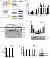
Fig 1
Fig 1. Overexpression of UBXN1 inhibits retrovirus production and NFκB pathway.
(A) Schematic of UBXN1 deletion and mutant constructs: yellow is N terminal UBA domain thought to bind polyubiquitin, green is coil-colied domain thought to mediate self-association, blue is UBX domain thought to interact with other proteins; (B) Quantification of HIV-eYFP (VSV G) production, after 293T co-transfection of HIV vector components and UBXN1 expression plasmid shown at bottom, with titer on HOS cells normalized to empty plasmid (set at 1), as measured by FACS; (C) Immunoblot of the various FLAG-UBXN1 mutant proteins, as detected using anti-FLAG antibody; (D) Quantification by FACS of HIV LTR activity in 293T cells transfected with decreasing amounts of 1–297 UBXN1 along with HIV-eYFP, as measured by eYFP MFI, normalized to empty vector (EV) (black bars); also quantification by RLU of HIV LTR-FFLUC, also normalized to EV (grey bars); underneath is shown immunoblot of transfected FLAG-tagged UBXN1 wt and AAA mutant proteins; (E) Quantification of transfection efficiency in 293T producers of other lentiviral vectors, as measured by eGFP/eYFP MFI by FACS in the presence of either FLAG-UBXN1 (grey bars) or EV (black bars); (F) As in part (B), quantification of production of indicated retroviral vectors, after co-transfection of FLAG-UBXN1 (grey bars), FLAG-AAA mutant (open bars), or EV (black bars) expression plasmids along with packaging and transfer vector components and VSV G into 293T cells, with resultant titer normalized to that of EV (set at 1.0 in each case). Shown to the right is a representative immunoblot of wt UBXN1 and AAA mutant. ****p < 0.0001, ***p < 0.0005, **p < 0.005, compared to EV, by two-way ANOVA.
Fig 2
Fig 2. Characterization of UBXN1 functional domains by reporter assay.
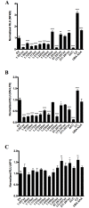
(A) Quantification of NFκB-FFLUC reporter in HEK293 cells 48 h after 96-well format transfection with indicated Myc-UBXN1 constructs (80 ng per well) or empty plasmid (Vector), in the presence 5 ng/mL TNFα for 4h; (B) Similar to (A), using an HIV LTR-FFLUC reporter; (C) Similar to (A), using an AP1-FFLUC reporter. All data represent mean ± SEM (n = 3). ****p < 0.0001, ***p < 0.0005, **p < 0.005, *p < 0.05, compared to vector alone, by two-way ANOVA.
Fig 3
Fig 3. UBXN1 interacts with Cullin1 and prevents IκBα degradation.
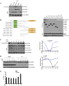
(A) FLAG-UBXN1 was co-transfected with Myc-Cul1, as indicated, and immunoprecipitated (IP) using anti-FLAG antibody, followed by immunoblotting (IB) using anti-FLAG or Myc antibody; β−tubulin served as loading control; (B) Left: schematic of Cul1 deletion constructs, with indicated regions that bind to Skp1 and Rbx1 (green and yellow, respectively, not precisely to scale); right: FLAG-Cul1 constructs were transfected into HEK293 cells, as indicated at top; endogenous UBXN1 was detected using anti-UBXN1 antibody and β−tubulin again served as loading control; (C) left: At top: Co-IP of endogenous UBXN1 and Cul1 from HFFs, after stimulating cells with 10 ng/mL TNFα; input of various proteins shown at bottom; right: relative band intensity of IκBα and Cul1, normalized against the density of β−tubulin (in the left immunoblot image); (D) left: Transfection of HEK293 cells with either empty vector or plasmid encoding FLAG-UBXN1, treated with 5 ng/mL TNFα for the indicated times, and cell lysates immunoblotted for all three indicated proteins, with β−tubulin serving as loading control; right: relative band intensity of IκBα normalized against the density of β−tubulin (in the left immunoblot image); (E) Similar to (D) except that NFκB-FFLUC reporter was co-transfected and cell lysates assayed for luciferase activity, normalized to co-transfected Renilla-LUC reporter, in the presence (grey bars) or absence (black bars) of UBXN1. Experiments of (C) and (D) were repeated at least twice, with similar results.
Fig 4
Fig 4. Co-immunoprecipitation of UBXN1, Rbx1, Skp1, and Cul1.
(A) 293T cells were transiently transfected with the indicated FLAG-UBXN1 constructs or FLAG-Cul1 in the presence of either Myc-Rbx1 or Myc-Skp1 as indicated, with co-IP at top and input, including β-tubulin, at bottom; (B) Similar to (A), except decreasing amounts of Myc-UBXN1 were transfected along with Myc-Skp1 and either empty vector (EV), ½-1 FLAG-Cul1, FLAG-Cul1, or 1/3-1 FLAG-Cul1, as indicated, with co-IP at top and input, including β-tubulin, at bottom; (C) Similar to (B), except that Myc-Rbx1 was transfected along with EV, either 0-1/2 FLAG-Cul1, FLAG-Cul1, or 0-2/3 FLAG-Cul1, as indicated, with co-IP at top and input, including Cox IV, at bottom. Note that co-expression of Cul1 increased Rbx1 levels.
Fig 5
Fig 5. Knockdown UBXN1 results in enhanced retroviral vector production and NFκB signaling.
(A) Quantification of HIV vector production after co-transfection of third-generation HIV-based vector encoding both anti-UBXN1 shRNA and puror gene (vs. control, empty vector), along with VSV G and either FG12, HIV-eYFP, or FG12-SV40 into 293T cells. FG12 is a third generation, self-inactivating HIV vector that encodes eGFP driven by the UbiC promote; FG12-SV40 is similar except it has an internal SV40 promoter driving eGFP. Top: puror titer on HOS targets; Bottom: percentage of eYFP/eGFP+ HOS targets as measured by flow cytometry for each of the three vectors, using two different amounts of indicated vector supernatant; (B) (left) Quantification of NFκB-FFLUC reporter in HEK293 cells after siRNA knockdown of UBXN1, compared to Trilencer-27 Universal scrambled negative control siRNA; (right) corresponding immunoblot of UBXN1 and β-tubulin in presence of either control or anti-UBXN1 siRNA. FFLUC values were normalized to those of co-transfected Renilla luciferase reporter; cells were stimulated for 4 h with 5 ng/mL of TNFα 48h post-transfection. (C) Similar to (B) except that either empty HIV vector pLK0.1 or vector encoding anti-UBXN1 shRNA was transfected into HEK293 cells, with corresponding immunoblot shown on right. Data represent mean ± SEM (n = 3). *p < 0.05, **p < 0.005, ***p < 0.0005, **** p<0.0001 by two-way ANOVA.
Fig 6
Fig 6. UBXN1 blocks NFκB signaling and inhibits HIV LTR activity.
(A) Left: immunoblot of indicated proteins from HFFs stably transduced with either empty HIV-based vector or vector encoding anti-UBXN1 shRNA, after stimulation with 5 ng/mL TNFα for indicated times; right: relative band intensity of IκBα, normalized to β−tubulin (in the left immunoblot image); (B) Quantification of NFκB (left) and HIV LTR (right) FFLUC reporters in cell lines of (A), normalized to co-transfected Renilla-LUC plasmid; for NFκB reporter cells were treated for 4 h with 5 ng/mL TNFα 48 h post-transfection; (C) Left: immunoblot of indicated proteins from UBXN1-/- HPRT-/- and control HPRT-/- MEFs, after stimulation with 10 ng/mL TNFα for indicated times (upper) or treated with 1 μM Bortezomib for 5 h and then stimulated with 10 ng/mL TNFα for the indicated times (lower); right: relative band intensity of IκBα, normalized to β−tubulin (in the left immunoblot images); (D) Confocal immunofluorescence microscopy of UBXN1 and Cul1 in UBXN1-/- HPRT-/- and control HPRT-/- MEFs. Nuclear DNA stained with TO-PRO-3 (I); UBXN1 (II) and Cul1 (III) were stained using secondary antibodies conjugated to Alexa Fluor 546 and 488, respectively; IV shows merge; Scale bar = 10 μm; (E) Quantification of NFκB and HIV LTR-FFLUC reporters in UBXN1-/- HPRT-/- and control HPRT-/- MEFs. Left: NFκB-FFLUC values 48 h post-transfection in the presence of 5 ng/mL TNFα for 4h, normalized to Renilla-LUC reporter; right: HIV LTR-FFLUC values 48 hrs post-transfection, similarly normalized; (F) JLAT10.6 T cells stably transduced with either HIV-based vector encoding anti-UBXN1 shRNA or control empty vector, after stimulating the cells with 5 ng/mL TNFα for the indicated times and measuring % eGFP+ by FACS. Data in (B) and (E) represent mean ± SEM (n = 3). **p < 0.005, *p < 0.05 by student’s t-test. Experiments of (A) and (C) were repeated several times, with similar results.
Fig 7
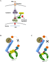
Fig 7. Schematic models of UBXN1 interaction with Cul1.
(A) Cartoon rendering of how UBXN1 may be interfering with Cul1 in the canonical NFκB signaling pathway; (B) and (C) additional schematics of how UBXN1 may somehow be interfering with Cul1 activity either by steric hindrance (B) or allosteric effect (C). UBXN1 is known to multimerize and interacts with both N and C termini of Cul1; stoichiometry between UBXN1 and Cul1 is not known (dashed ovals). In both models Skp1 and Rbx1 interact with the N and C termini of Cul1, respectively, and UBXN1 interacts with Rbx1 but not Skp1. These models do not exclude the possibility that another factor or protein (e.g., multimerized ubiquitin) mediates the interaction between UBXN1 and Cul1/Rbx1.

Similar articles

Cited by

References

    1. Kano F., Kondo H., Yamamoto A., Tanaka A.R., Hosokawa N., Nagata K., and Murata M. The maintenance of the endoplasmic reticulum network is regulated by p47, a cofactor of p97, through phosphorylation by cdc2 kinase. Genes Cells 2005; 10, 333–344. 10.1111/j.1365-2443.2005.00837.x - DOI - PubMed
    1. Kondo H., Rabouille C., Newman R., Levine T.P., Pappin D., Freemont P., and Warren G. p47 is a cofactor for p97-mediated membrane fusion. Nature 1997; 388, 75–78. 10.1038/40411 - DOI - PubMed
    1. Meyer H.H., Kondo H., and Warren G. The p47 co-factor regulates the ATPase activity of the membrane fusion protein, p97. FEBS Lett 1998; 437, 255–257. - PubMed
    1. Uchiyama K., Jokitalo E., Lindman M., Jackman M., Kano F., Murata M., Zhang X., and Kondo H. The localization and phosphorylation of p47 are important for Golgi disassembly-assembly during the cell cycle. J Cell Biol 2003; 161, 1067–1079. 10.1083/jcb.200303048 - DOI - PMC - PubMed
    1. Uchiyama K., and Kondo H. p97/p47-Mediated biogenesis of Golgi and ER. J Biochem 2005; 137, 115–119. 10.1093/jb/mvi028 - DOI - PubMed

Publication types