. 2017 Mar 14;116(6):775-784.
doi: 10.1038/bjc.2017.15. Epub 2017 Feb 2.

Asporin is a stromally expressed marker associated with prostate cancer progression

Affiliations

Asporin is a stromally expressed marker associated with prostate cancer progression

Annie Rochette et al. Br J Cancer. .

Abstract

Background: Prostate cancer shows considerable heterogeneity in disease progression and we propose that markers expressed in tumour stroma may be reliable predictors of aggressive tumour subtypes.

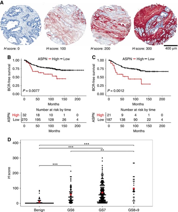
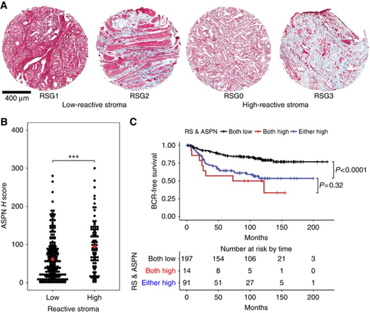
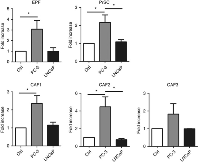
Methods: We have used Kaplan-Meier, univariate and multivariate analysis to correlate the expression of Asporin (ASPN) mRNA and protein with prostate cancer progression in independent cohorts. We used immunohistochemistry and H scoring to document stromal localisation of ASPN in a tissue microarray and mouse prostate cancer model, and correlated expression with reactive stroma, defined using Masson Trichrome staining. We used cell cultures of primary prostate cancer fibroblasts treated with serum-free conditioned media from prostate cancer cell lines to examine regulation of ASPN mRNA in tumour stromal cells.

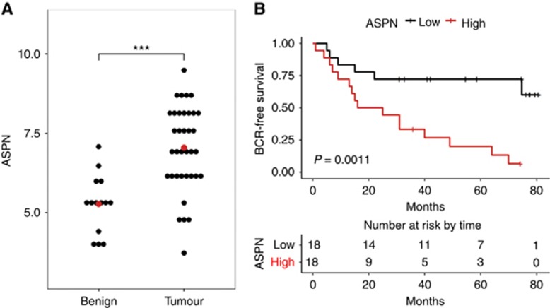
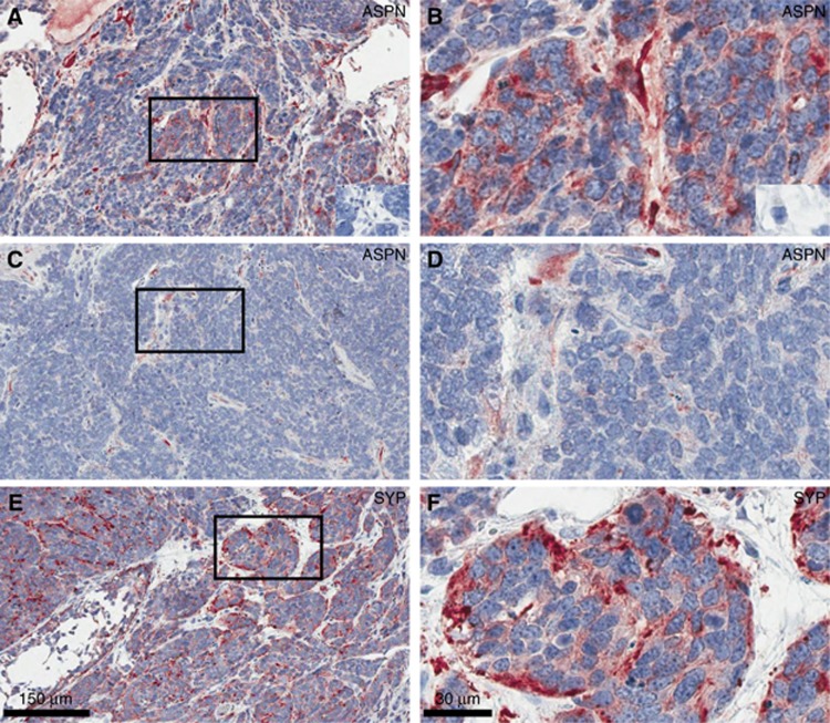
Results: We observed increased expression of ASPN mRNA in a data set derived from benign vs tumour microdissected tissue, and a correlation with biochemical recurrence using Kaplan-Meier and Cox proportional hazard analysis. ASPN protein localised to tumour stroma and elevated expression of ASPN was correlated with decreased time to biochemical recurrence, in a cohort of 326 patients with a median follow up of 9.6 years. Univariate and multivariate analysis demonstrated that ASPN was correlated with progression, as were Gleason score, and clinical stage. Additionally, ASPN expression correlated with the presence of reactive stroma, suggesting that it may be a stromal marker expressed in response to the presence of tumour cells and particularly with aggressive tumour subtypes. We observed expression of ASPN in the stroma of tumours induced by p53 inhibition in a mouse model of prostate cancer, and correlation with neuroendocrine marker expression. Finally, we demonstrated that ASPN transcript expression in normal and cancer fibroblasts was regulated by conditioned media derived from the PC3, but not LNCaP, prostate cancer cell lines.

Conclusions: Our results suggest that ASPN is a stromally expressed biomarker that correlates with disease progression, and is observed in reactive stroma. ASPN expression in stroma may be part of a stromal response to aggressive tumour subtypes.


Conflict of interest statement

The authors declare no conflict of interest.

Figures

Figure 1
Figure 1
Correlation of ASPN mRNA with BCR in the prostate cancer patient cohort described by Mortensen et al (2015). (A) A significant increase in ASPN transcripts was observed in tumour vs patient matched benign samples; mean ASPN mRNA levels of 7.0 in tumour samples vs 5.3 in benign samples (t-test, P<0.001). (B) Kaplan–Meier analysis correlated higher ASPN transcript levels with shorter time to biochemical recurrence (P=0.0011) in the Mortensen et al (2015) data set. The lower panel shows the number of patients at risk of BCR during 80 months of follow up.
Figure 2
Figure 2
Correlation of ASPN protein expression with BCR using IHC staining of a TMA with patient outcome data. Immunohistochemistry staining of ASPN was undertaken on the McGill cohort of 326 patients with follow up; outcome data and clinico-pathological details are listed in Supplementary Table S1. Asporin staining (brown/red) was observed in stroma and showed peri-tumoural distribution with varying intensity and extent. (A) Representative images of ASPN staining quantified by H-score, with examples of H-scores 0, 100, 200, and 300. Scale bar is 400 μm. H-score combines the staining extent and intensity observed, and was scored by two individuals. (B) Kaplan–Meier analysis of ASPN H-score demonstrated that patients with an H-score over 150 showed increased BCR (P=0.0077). (C) High ASPN (H-score>150) was correlated with increased progression (BCR) in the subset of patients with Gleason grade 7 (P=0.0012) relative to patients with low ASPN (H-score⩽150). Panel D: H-score values for ASPN staining and Gleason grade. There was low ASPN staining in benign tissue, and increased ASPN staining in Gleason grades 6, 7, and 8+9 (ANOVA followed by Tukey multiple comparison, *P<0.05, **P<0.01 and ***P<0.001). Mean ASPN levels in benign H-score=9.5, Gleason 6 H-score=55.9, Gleason 7 H-score=71.0, Gleason 8+9 H-score=97.4.
Figure 3
Figure 3
Co-identification of ASPN and reactive stromal histology. After IHC staining with ASPN and image storage, we stripped the TMA and performed Masson's trichrome staining to identify areas of reactive stroma. Low reactive stroma was scored as RSG1 and 2 while high reactive stroma was RSG0 and RSG3 (Ayala et al, 2003; Yanagisawa et al, 2007). (A) Representative images of samples graded as reactive stroma 1, 2, (low) and 0, and 3 (high) using Masson's trichrome. (B) Graph of patients showing ASPN staining and reactive stroma; ASPN staining was observed in patients with or without reactive stroma and there was evidence of elevated ASPN staining (H-score) and high reactive stroma (Low reactive stroma samples had a mean H-score of 61.5 while high reactive stroma samples showed an ASPN H-score of 94.3, t-test P=0.001). (C) Kaplan–Meier curve of progression among patients with no reactive stroma and low ASPN H-score (black), high reactive stroma and high ASPN (red), and patients showing either reactive stroma or ASPN (blue). Comparison among the three groups showed that samples positive for both reactive stroma and ASPN was predictive of progression as well as samples showing either high reactive stroma or ASPN (P<0.0001). There was no statistically significant difference between the double positive and single positive samples (P=0.32).
Figure 4
Figure 4
Expression of ASPN in the murine TP53 prostate cancer model. Asporin was localised by immunohistochemistry in the mouse probasin driven TP53 prostate cancer model to determine whether there was expression in stromal cells and/or tumour cells. Prostate tumours from prostate specific p53 knockout mice showed ASPN staining in the stromal compartment (low magnification, A and C; high magnification, B and D). Tumours showed few stromal cells, though many of those present were positive for ASPN. IHC controls without primary antibody are insert in A and B. Left panels × 10 magnification and right panels at × 40 magnification. Synaptophysin staining is shown in E and F.
Figure 5
Figure 5
The effect of conditioned media from prostate cancer cell lines upon ASPN expression levels in fibroblasts. Serum-free conditioned media was prepared from PC-3 and LNCaP cells, and added to fetal prostate fibroblasts (EPF), primary prostate stromal cells (PrSC), and 3 isolates of CAFs for 24 h followed by qRT–PCR analysis of ASPN mRNA. Fold increase compared to control media, and normalised to TBP internal control mRNA. Error bars indicate s.e.m. CM from PC-3 cells induced an increase of ASPN mRNA but LNCaP CM did not; in EPF, PrSC and 2 of 3 CAFs. (ANOVA followed by Tukey multiple comparison, *P<0.05).

References

    1. Andersen S, Solstad O, Moi L, Donnem T, Eilertsen M, Nordby Y, Ness N, Richardsen E, Busund LT, Bremnes RM (2015) Organized metabolic crime in prostate cancer: The coexpression of MCT1 in tumor and MCT4 in stroma is an independent prognosticator for biochemical failure. Urol Oncol 33: 338.e339–317. - PubMed
    1. Awata T, Yamada S, Tsushima K, Sakashita H, Yamaba S, Kajikawa T, Yamashita M, Takedachi M, Yanagita M, Kitamura M, Murakami S (2015) PLAP-1/Asporin Positively Regulates FGF-2 Activity. J Dent Res 94: 1417–1424. - PubMed
    1. Ayala G, Tuxhorn JA, Wheeler TM, Frolov A, Scardino PT, Ohori M, Wheeler M, Spitler J, Rowley DR (2003) Reactive stroma as a predictor of biochemical-free recurrence in prostate cancer. Clin Cancer Res 9: 4792–4801. - PubMed
    1. Bosman FT, de Goeij AF, Rousch M (1992) Quality control in immunocytochemistry: experiences with the oestrogen receptor assay. J Clin Pathol 45: 120–124. - PMC - PubMed
    1. Dakhova O, Ozen M, Creighton CJ, Li R, Ayala G, Rowley D, Ittmann M (2009) Global gene expression analysis of reactive stroma in prostate cancer. Clin Cancer Res 15: 3979–3989. - PMC - PubMed

MeSH terms