. 2017 Jun;23(6):813-825.
doi: 10.1002/lt.24735.

Phosphatase and tensin homolog-β-catenin signaling modulates regulatory T cells and inflammatory responses in mouse liver ischemia/reperfusion injury

Affiliations

Phosphatase and tensin homolog-β-catenin signaling modulates regulatory T cells and inflammatory responses in mouse liver ischemia/reperfusion injury

Qiang Zhu et al. Liver Transpl. 2017 Jun.

Abstract

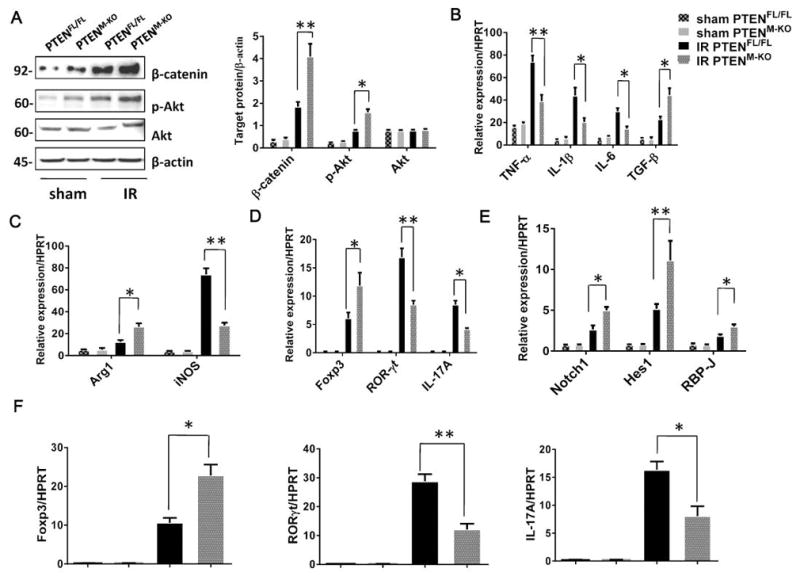
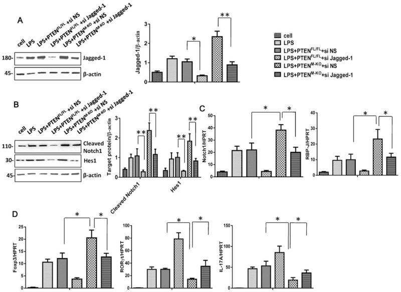
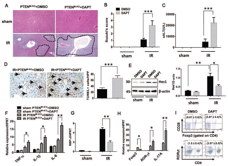
The phosphatase and tensin homolog (PTEN) deleted on chromosome 10 plays an important role in regulating T cell activation during inflammatory response. Activation of β-catenin is crucial for maintaining immune homeostasis. This study investigates the functional roles and molecular mechanisms by which PTEN-β-catenin signaling promotes regulatory T cell (Treg) induction in a mouse model of liver ischemia/reperfusion injury (IRI). We found that mice with myeloid-specific phosphatase and tensin homolog knockout (PTENM-KO ) exhibited reduced liver damage as evidenced by decreased levels of serum alanine aminotransferase, intrahepatic macrophage trafficking, and proinflammatory mediators compared with the PTEN-proficient (floxed phosphatase and tensin homolog [PTENFL/FL ]) controls. Disruption of myeloid PTEN-activated b-catenin promoted peroxisome proliferator-activated receptor gamma (PPARγ)-mediated Jagged-1/Notch signaling and induced forkhead box P3 (FOXP3)1 Tregs while inhibiting T helper 17 cells. However, blocking of Notch signaling by inhibiting γ-secretase reversed myeloid PTEN deficiency-mediated protection in ischemia/reperfusion-triggered liver inflammation with reduced FOXP3+ and increased retinoid A receptor-related orphan receptor gamma t-mediated interleukin 17A expression in ischemic livers. Moreover, knockdown of β-catenin or PPARγ in PTEN-deficient macrophages inhibited Jagged-1/Notch activation and reduced FOXP3+ Treg induction, leading to increased proinflammatory mediators in macrophage/T cell cocultures. In conclusion, our findings demonstrate that PTEN-β-catenin signaling is a novel regulator involved in modulating Treg development and provides a potential therapeutic target in liver IRI. Liver Transplantation 23 813-825 2017 AASLD.


Conflict of interest statement

The authors declare no conflicts of interest.

Figures

Figure 1
Figure 1. Myeloid PTEN deficiency ameliorates hepatocellular damage and reduces macrophage trafficking in IR-induced liver injury
(A) Representative histological staining (H&E) of ischemic liver tissue (n=4–5/group, magnification x100). (B) Liver damage, evaluated by Suzuki’s score. ***p<0.001. (C) Hepatocellular function, assessed by serum ALT levels (IU/L). Results expressed as mean±SD (n=4–5/group), ***p<0.001. (D) Liver apoptosis by TUNEL staining. Results expressed as mean±SD (n=4–6/group, magnification x400), ***p<0.001. (E) Immunofluorescence staining of CD68+ macrophages (short arrow) in ischemic liver lobes. Results expressed as mean±SD (n=4/group, magnification ×400), ***p<0.001.
Figure 2
Figure 2. Myeloid PTEN deficiency promotes β-catenin activation and Treg induction in IR-induced liver injury
(A) Western blot analysis and relative density ratio of β-catenin, p-Akt, and Akt in ischemic livers. *p<0.05, **p<0.01. Quantitative RT-PCR-assisted detection of (B) TNF-α, IL-1β, IL-6, TGF-β, (C) Arg1, iNOS, and (D) Foxp3, RORγt, IL-17A in ischemic livers or (E) Notch1, Hes1, RBP-J and (F) Foxp3, RORγt, IL-17A in spleen T cells from PTENFL/FL and PTENM-KO mice. Each column represents the mean±SD (n=3–4/group). *p<0.05, **p<0.01.
Figure 3
Figure 3. PTEN-β-catenin axis activates PPARγ and Jagged-1/Notch signaling pathway and induces Foxp3+Tregs in vitro
BMMs were transfected with β-catenin siRNA (siβ-cat), and then co-cultured with spleen T cells after LPS stimulation for 6 h. (A) Western blot analysis and relative density ratio of β-catenin, PPARγ and Jagged-1 in LPS-stimulated macrophages. *p<0.05, **p<0.01. Quantitative RT-PCR-assisted detection of (B) TNF-α, IL-1β, IL-6, TGF-β, (C) Arg1, iNOS in LPS-stimulated macrophages. Each column represents the mean±SD (n=3–4/group). *p<0.05, **p<0.01. (D) Western blot analysis and relative density ratio of cleaved Notch1 in spleen T cells after co-culture *p<0.05, **p<0.01. Quantitative RT-PCR-assisted detection of (E) Notch1, Hes1, RBP-J and (F) Foxp3, RORγt, IL-17A in spleen T cells after co-culture. Each column represents the mean±SD (n=3–4/group). *p<0.05, **p<0.01.
Figure 4
Figure 4. PPARγ mediates Jagged-1/Notch signaling pathway in vitro
BMMs were transfected with PPARγ siRNA (siPPARγ), and then co-cultured with spleen T cells after LPS stimulation for 6 h. (A) Western blot analysis and relative density ratio of PPARγ and Jagged-1 in LPS-stimulated macrophages. *p<0.05, **p<0.01. Quantitative RT-PCR-assisted detection of (B) TNF-α, IL-1β, IL-6, TGF-β, (C) Arg1, iNOS in LPS-stimulated macrophages. Each column represents the mean±SD (n=3–4/group). *p<0.05, **p<0.01. (D) Western blot analysis and relative density ratio of cleaved Notch1 in spleen T cells after co-culture *p<0.05. Quantitative RT-PCR-assisted detection of (E) Notch1, RBP-J and (F) Foxp3, RORγt, IL-17A in spleen T cells after co-culture. Each column represents the mean±SD (n=3–4/group). *p<0.05, **p<0.01.
Figure 5
Figure 5. Jagged-1/Notch signaling is essential for the Foxp3+Treg induction in the PTEN-β-catenin signaling-mediated immune regulation in vitro
BMMs were transfected with Jagged-1 siRNA (siJagged-1), and then co-cultured with spleen T cells after LPS stimulation for 6 h. (A) Western blot analysis and relative density ratio of Jagged-1 in LPS-stimulated macrophages. *p<0.05, **p<0.01. Quantitative RT-PCR-assisted detection of (B) Western blot analysis and relative density ratio of cleaved Notch1 in spleen T cells after co-culture *p<0.05, **p<0.01. Quantitative RT-PCR-assisted detection of (C) Notch1, RBP-J and (D) Foxp3, RORγt, IL-17A in spleen T cells after co-culture. Each column represents the mean±SD (n=3–4/group). *p<0.05.
Figure 6
Figure 6. Blocking Jagged-1/Notch signaling pathway aggravates IR-induced liver damage and inhibits Foxp3+Treg induction in vivo
(A) Representative histological staining (H&E) of ischemic livers. (n=4–5/group, magnification x100). (B) The severity of liver IRI was evaluated by the Suzuki’s histological grading. ***p<0.001. (C) Hepatocellular function was evaluated by sALT levels (IU/L). Results expressed as mean±SD (n=4–5/group). ***p<0.001. (D) Liver apoptosis by TUNEL staining. Results expressed as mean±SD (n=4–6/group, magnification x400), ***p<0.001. (E) Western blot analysis and relative density ratio of Hes1 in ischemic livers. *p<0.05, **p<0.01. Quantitative RT-PCR-assisted detection of (F) TNF-α, IL-1β, IL-6, (G) RBP-J, and (H) Foxp3, RORγt, IL-17A in ischemic livers. Each column represents the mean±SD (n=3–4/group). *p<0.05, **p<0.01. (I) Foxp3 and RORγt expression in spleen T cells were evaluated by flow cytometry. Results expressed as mean±SD (n=3/group, p<0.001).
Figure 7
Figure 7
Schematic illustration of molecular mechanisms of PTEN-β-catenin axis in the regulation of regulatory T cells and inflammatory responses in liver IRI.

References

    1. Peralta C, Jimenez-Castro MB, Gracia-Sancho J. Hepatic ischemia and reperfusion injury: effects on the liver sinusoidal milieu. Journal of hepatology. 2013;59(5):1094–106. - PubMed
    1. Ju C, Tacke F. Hepatic macrophages in homeostasis and liver diseases: from pathogenesis to novel therapeutic strategies. Cellular & molecular immunology. 2016 - PMC - PubMed
    1. Colletti LM, Remick DG, Burtch GD, Kunkel SL, Strieter RM, Campbell DA., Jr Role of tumor necrosis factor-alpha in the pathophysiologic alterations after hepatic ischemia/reperfusion injury in the rat. The Journal of clinical investigation. 1990;85(6):1936–43. - PMC - PubMed
    1. Zwacka RM, Zhang Y, Halldorson J, Schlossberg H, Dudus L, Engelhardt JF. CD4(+) T-lymphocytes mediate ischemia/reperfusion-induced inflammatory responses in mouse liver. The Journal of clinical investigation. 1997;100(2):279–89. - PMC - PubMed
    1. Eggenhofer E, Rovira J, Sabet-Baktach M, Groell A, Scherer MN, Dahlke MH, et al. Unconventional RORgammat+ T cells drive hepatic ischemia reperfusion injury. Journal of immunology. 2013;191(1):480–7. - PubMed

Publication types

MeSH terms