Review
. 2017 May:58:45-69.
doi: 10.1016/j.preteyeres.2017.01.006. Epub 2017 Jan 30.

Bestrophin 1 and retinal disease

Affiliations
Review

Bestrophin 1 and retinal disease

Adiv A Johnson et al. Prog Retin Eye Res. 2017 May.

Abstract

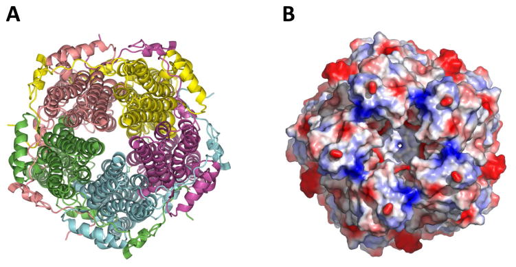
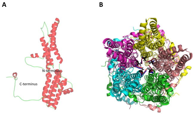
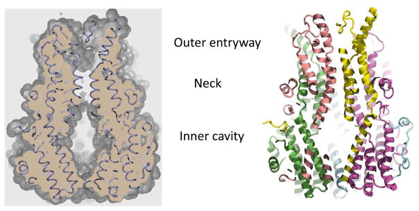
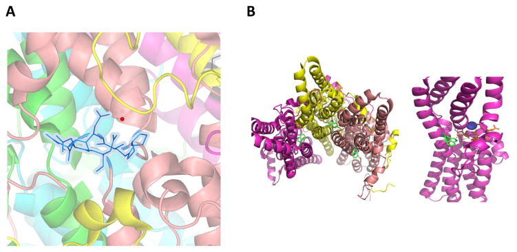
Mutations in the gene BEST1 are causally associated with as many as five clinically distinct retinal degenerative diseases, which are collectively referred to as the "bestrophinopathies". These five associated diseases are: Best vitelliform macular dystrophy, autosomal recessive bestrophinopathy, adult-onset vitelliform macular dystrophy, autosomal dominant vitreoretinochoroidopathy, and retinitis pigmentosa. The most common of these is Best vitelliform macular dystrophy. Bestrophin 1 (Best1), the protein encoded by the gene BEST1, has been the subject of a great deal of research since it was first identified nearly two decades ago. Today we know that Best1 functions as both a pentameric anion channel and a regulator of intracellular Ca2+ signaling. Best1 is an integral membrane protein which, within the eye, is uniquely expressed in the retinal pigment epithelium where it predominantly localizes to the basolateral plasma membrane. Within the brain, Best1 expression has been documented in both glial cells and astrocytes where it functions in both tonic GABA release and glutamate transport. The crystal structure of Best1 has revealed critical information about how Best1 functions as an ion channel and how Ca2+ regulates that function. Studies using animal models have led to critical insights into the physiological roles of Best1 and advances in stem cell technology have allowed for the development of patient-derived, "disease in a dish" models. In this article we review our knowledge of Best1 and discuss prospects for near-term clinical trials to test therapies for the bestrophinopathies, a currently incurable and untreatable set of diseases.

Keywords: Anion channel; Best1; Bestrophin; Maculopathy; Retinal disease; Retinal pigment epithelium.


Figures

Figure 1
Figure 1. Clinical presentation of Best vitelliform macular dystrophy
A classic vitelliform lesion is found in both the right (A) and left (D) eye of an 80 year old, female patient with Best disease. She presented with mild hyperopia, 20/40 vision in one eye, and 20/400 vision in the other eye. Both lesions were autofluorescent (B, E). OCT imaging of a horizontal section of the left (C) and right (F) maculas revealed retinal abnormalities. In particular, the left eye showed a raised retina and multiple fluid-filled, serous retinal detachments.
Figure 2
Figure 2. Clinical presentation of multifocal Best vitelliform macular dystrophy
Fundus photographs revealed prominent multi-focal lesions in both the left (A, C) and right (B, E) eyes in a 33 year-old male patient with multifocal Best disease. These lesions were autofluorescent (D, F) and choroidal neovascularization was apparent in the fundus of the patient’s left eye (E).
Figure 3
Figure 3. Clinical presentation of adult-onset vitelliform macular dystrophy
While initially thought to have age-related macular degeneration, further testing diagnosed this 88 year old female patient with adult-onset vitelliform macular dystrophy. The presentation is identical to Best vitelliform macular dystrophy, with a classical vitelliform lesion in the fundus of both eyes (A, D). These lesions are autofluorescent (B, E) and OCT imaging of a horizontal section of the macula shows retinal abnormalities (C, F). The OCT of the left eye, in particular, shows a fluid-filled retinal detachment (C).
Figure 4
Figure 4. Clinical presentation of autosomal recessive bestrophinopathy
Fundus photographs of a 17-year old girl diagnosed with autosomal recessive bestrophinopathy show classical findings, such as vitelliform lesions (A–F) and yellowish, subretinal deposits (G, H). These lesions are autofluorescent (I).
Figure 5
Figure 5. Clinical presentation of autosomal dominant vitreoretinochoroidopathy
Fundus photographs of a patient diagnosed with autosomal dominant vitreoretinochoroidopathy reveal classical symptoms, including a sharp demarcation line between a region of normal retina and a region of clumped, hyperpigmentation. Whitish specs and yellowish deposits are distributed throughout the peripheral retina.
Figure 6
Figure 6. Basolateral plasma membrane localization of Best1 in confluent MDCK II, fhRPE, and iPSC-RPE cells
Best1 was expressed in MDCK II cells via adenovirus mediated gene transfer and stained for Best1 (green) and the apical plasma membrane marker Gp135 (red). Localization of endogenous Best1 (green) was assessed in both fhRPE and iPSC-RPE cells. For both fhRPE and iPSC-RPE cells, nuclei (blue) were stained as a positional marker. iPSC-RPE cells were additionally stained with the tight junction marker ZO-1 (red). Confocal X-Y and X-Z scans were generated to show the localization of Best1 in the X, Y, and Z planes for all three cell types. Scale bars: 20 μm.
Figure 7
Figure 7. Neuronal expression of localization of mBest1
A) Western blot analysis of mBest1 in cultured astrocytes and gene silencing for mBest1 by infection with lentivirus carrying mB1-shRNA or control-shRNA. B) Immunostaining of DAPI (blue), GFAP (green), and mBest1 (red) of hippocampal CA1 region in wild-type mouse (left) and Best1 KO mouse (right). Best1 is strongly co-localized with GFAP, an astrocytic marker, in wild-type mice, while mBest1 is not expressed in mBest1 KO mice. C) Immunostaining for mBest1 (red) and GFP (green) in cerebellum from GFAP-GFP transgenic mice. mBest1 is expressed in Purkinje cells (asterisk), interneurons (white arrowheads), Bergmann glia (arrows), and lamellar astrocytes (pale blue arrowheads), but not in granule cells. All GFP-positive astrocytes robustly expressed mBest1. D) Immunostaining and quantification of Best1 in the molecular layer of DG in mouse hippocampus. Top, representative confocal images for mBest1 (red) and GFP (green) in astrocytes. Bottom, percentage of Best1-positive areas in the cell body and process or in the microdomain over total area. **P < 0.01 (Student’s t-test). E) Immunogold electron microscopy of Best1 in the molecular layer of DG in mouse hippocampus. Top, representative images of mBest1 labeling (black dots indicated by arrowheads) in DAB-stained astrocytes (outlined with dashed lines). Pre, presynaptic terminal; Post, postsynapse. Bottom left, density of gold particles for Best1 in cell body, process and microdomain. Bottom right, percentage of gold particles for Best1 located on the plasma membrane of the cell body, process, and microdomain. ***P < 0.001 (Student’s t-test). Number on each bar refers to the number of cells (d) or images (e) analyzed. Data are presented as mean ± s.e.m.
Figure 8
Figure 8. Model diagram of memory impairment in Alzheimer’s disease
In Alzheimer’s disease, astrocytes near Amyloid β plaques (a) have more putrescine (b). Putrescine is degraded by MAOB (c) to produce the inhibitory neurotransmitter GABA (d). GABA is then abnormally released via BEST1 channels (e) which is redistributed away from microdomains. The released GABA binds to extrasynaptic GABAA and GABAB receptors (f) and strongly inhibits presynaptic release and spike probability. Consequently, granule cells of the dentate gyrus receive less glutamatergic inputs at perforant path synapses and show reduced synaptic plasticity. This finally leads to memory impairment in Alzheimer disease. Pre: presynaptic terminal, Post: postsynapse, NMDAR: N-methyl-D-aspartate receptor, AMPAR: α-amino-3-hydroxy-5-methyl-4-isoxazole propionate receptor.
Figure 9
Figure 9. Best1 forms homo-pentameric anion channels
A) Cartoon representation showing the pentameric arrangement of the subunits of the Best1 channel. B) A detailed surface representation of the Best1 channel from the top, showing the opening of the pore formed from five identical subunits.
Figure 10
Figure 10. The Best1 protomer and neck region of the pore
A) Ribbon diagram of a Best1 protomer. B) Cartoon representation of the pentameric channel. View from top, showing the amino acids which form the first restriction site in the neck region of the pore. Ile 76 in black, Phe 80 in red, and Phe 84 in blue.
Figure 11
Figure 11. A cut-away view of Best1 showing the pore
The outer entryway, neck, and inner cavity are the three major compartments that comprise the pore.
Figure 12
Figure 12. Best1 pentameric channels contain a calcium clasp region
A) The view of the calcium clasp site. Both calcium (red sphere) and the amino acid side chains (blue) involved in the coordination of Ca2+ are highlighted. The amino acids are Pro297, Glu 300, Asp 301–304. B) Left. The view of location of Trp 93. Trp is not part of the restriction of the pore. Right. Trp 93 is located closer to the Ca2+ clasp region.
Figure 13
Figure 13. Clinical manifestation of canine bestrophinopathy
(A) Fundus photograph of a 23-week-old cBest1-R25X-affected dog with focal canine macular lesion in vitelliform stage (arrowhead). (B) A 36-week-old cBest1-R25X/P463fs-affected dog exhibiting early stage lesions associated with both canine fovea-like region (arrowhead) and aligned along visual streak. (C) Multifocal presentation of canine bestrophinopathy in 40-week-old dog harboring cBest1-R25X/P463fs compound heterozygous mutation. Arrowhead indicates lesion in fovea-like region. OD: right eye; OS: left eye; cBest1: canine bestrophin 1.
Figure 14
Figure 14. Progression of unifocal disease in canine bestrophinopathy
(A) cSLO/SD-OCT images of central lesion in previtelliform stage (OD) in a 15-week-old cBest1-R25X-affected dog. Note the subtle dissociation of the neural retina from the RPE on the OCT scan (arrowhead). (B) En face infrared view of representative cBEST1-C73T/R25X mutant dog. (*) indicates canine fovea-like area in OS; black arrows signify locations of cross-sectional OCT scans shown in the below panel. Outer photoreceptor nuclear layer (ONL) and retinal pigment epithelium (RPE) are highlighted for visibility on OCT scans. (C) Topographic localization of the sites (*) of the early macular lesions in cBest1-mutant dogs (ages: 10–62 weeks; n = 7, left) correspond to the localization of the fovea-like area in wildtype dogs (ages: 7 weeks – 8 years; n = 13, right). (D) A 17-week-old cBest1-R25X-affected dog with a classic circular vitelliform lesion (OD) resembling Stage II of Best Vitelliform Macular Dystrophy (BVMD). OD: right eye; OS: left eye; NIR - near infrared reflectance. Images B & C taken from Beltran et al, 2014 (doi:10.1371/journal.pone.0090390.g002).
Figure 15
Figure 15. Immunohistochemical evaluation of an early lesion in the canine model of Best1-associated maculopathies
A vitelliform lesion of a 112-week-old cBest1-R25X-affected dog immunolabeled with anti-cone arrestin (hCAR, red). Note the massive autofluorescent deposits within RPE cell monolayer and in the subretinal space (green and yellowish-green: native autofluorescence). RPE: retinal pigment epithelium; PRs: photoreceptors. ONL: outer nuclear layer; INL: inner nuclear layer. Scale bar 40 μm.
Figure 16
Figure 16. Comparison of Best1+/+ and Best1 knock-in mice
Fundus from a wild type mouse (A) and a Best1 knock-in mouse harboring the disease-causing mutation W93C (B). The dotted line highlights the anomalous portion of the fundus in the Best1 knock-in mouse (B). Panel C shows an electron micrograph of a chronic serous detachment of the retina (*) in a Best1 knock-in mouse harboring the disease-causing mutation W93C. Note the RPE microvilli lying horizontally (mv). Photoreceptor outer segments show damage and internal debris (arrows). Panel D shows that, in some regions, unphagocytosed photoreceptor outer segments (arrowheads) are observed “sitting” atop RPE microvilli. Note the extensive accumulation of lipofuscin granules in both C and D.

References

    1. Abramoff MD, Mullins RF, Lee K, Hoffmann JM, Sonka M, Critser DB, Stasheff SF, Stone EM. Human photoreceptor outer segments shorten during light adaptation. Investigative ophthalmology & visual science. 2013;54:3721–3728. - PMC - PubMed
    1. Al-Jumaily M, Kozlenkov A, Mechaly I, Fichard A, Matha V, Scamps F, Valmier J, Carroll P. Expression of three distinct families of calcium-activated chloride channel genes in the mouse dorsal root ganglion. Neuroscience bulletin. 2007;23:293–299. - PMC - PubMed
    1. Allikmets R, Seddon JM, Bernstein PS, Hutchinson A, Atkinson A, Sharma S, Gerrard B, Li W, Metzker ML, Wadelius C, Caskey CT, Dean M, Petrukhin K. Evaluation of the Best disease gene in patients with age-related macular degeneration and other maculopathies. Human genetics. 1999;104:449–453. - PubMed
    1. Andre S, Boukhaddaoui H, Campo B, Al-Jumaily M, Mayeux V, Greuet D, Valmier J, Scamps F. Axotomy-induced expression of calcium-activated chloride current in subpopulations of mouse dorsal root ganglion neurons. Journal of neurophysiology. 2003;90:3764–3773. - PubMed
    1. Arndt CF, Derambure P, Defoort-Dhellemmes S, Hache JC. Outer retinal dysfunction in patients treated with vigabatrin. Neurology. 1999;52:1201–1205. - PubMed

Publication types