. 2017 Mar 10;292(10):4210-4221.
doi: 10.1074/jbc.M116.767897. Epub 2017 Feb 2.

Extracellular Loops Are Essential for the Assembly and Function of Polycystin Receptor-Ion Channel Complexes

Affiliations

Extracellular Loops Are Essential for the Assembly and Function of Polycystin Receptor-Ion Channel Complexes

Zahra Salehi-Najafabadi et al. J Biol Chem. .

Abstract

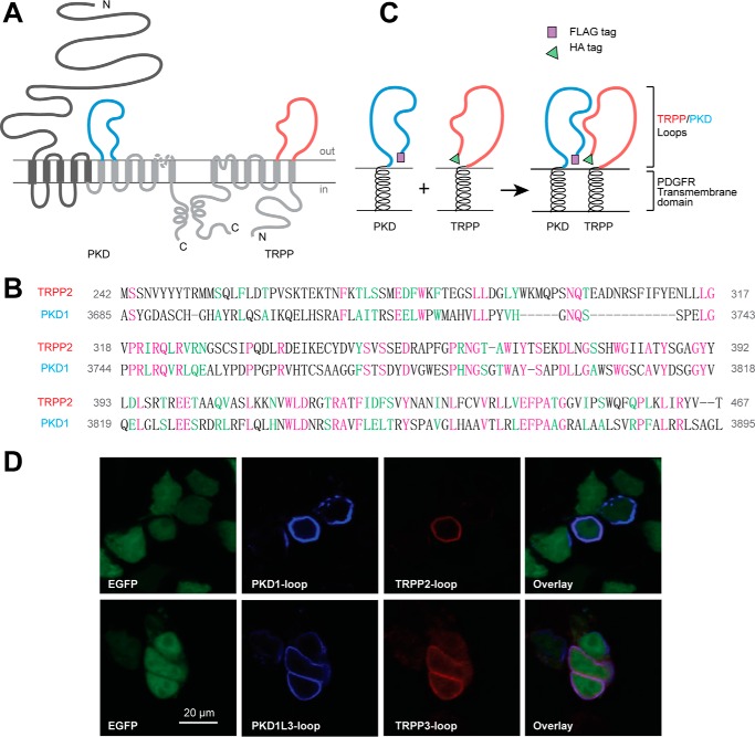
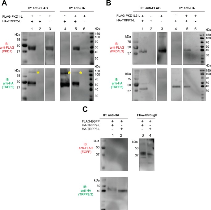
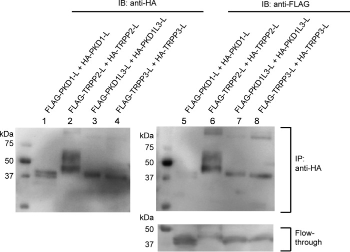
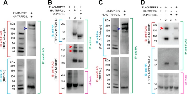
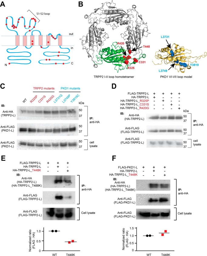
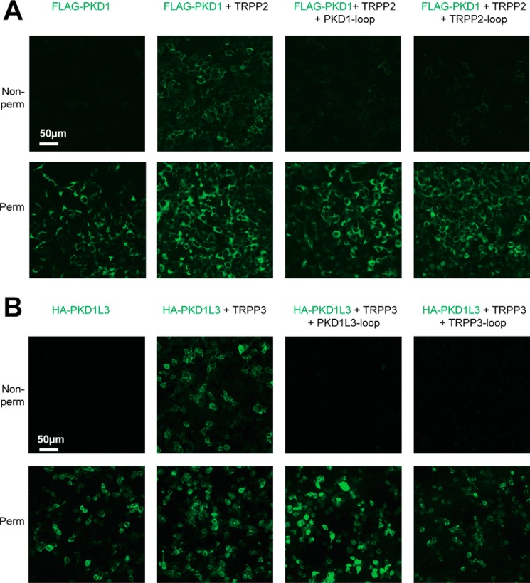
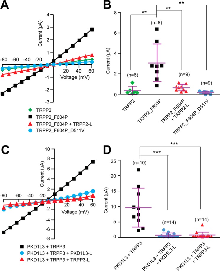
Polycystin complexes, or TRPP-PKD complexes, made of transient receptor potential channel polycystin (TRPP) and polycystic kidney disease (PKD) proteins, play key roles in coupling extracellular stimuli with intracellular Ca2+ signals. For example, the TRPP2-PKD1 complex has a crucial function in renal physiology, with mutations in either protein causing autosomal dominant polycystic kidney disease. In contrast, the TRPP3-PKD1L3 complex responds to low pH and was proposed to be a sour taste receptor candidate. It has been shown previously that the protein partners interact via association of the C-terminal or transmembrane segments, with consequences for the assembly, surface expression, and function of the polycystin complexes. However, the roles of extracellular components, especially the loops that connect the transmembrane segments, in the assembly and function of the polycystin complex are largely unknown. Here, with an immunoprecipitation method, we found that extracellular loops between the first and second transmembrane segments of TRPP2 and TRPP3 associate with the extracellular loops between the sixth and seventh transmembrane segments of PKD1 and PKD1L3, respectively. Immunofluorescence and electrophysiology data further confirm that the associations between these loops are essential for the trafficking and function of the complexes. Interestingly, most of the extracellular loops are also found to be involved in homomeric assembly. Furthermore, autosomal dominant polycystic kidney disease-associated TRPP2 mutant T448K significantly weakened TRPP2 homomeric assembly but had no obvious effect on TRPP2-PKD1 heteromeric assembly. Our results demonstrate a crucial role of these functionally underexplored extracellular loops in the assembly and function of the polycystin complexes.

Keywords: PKD; TRPP; autosomal dominant polycystic kidney disease; cell surface receptor; extracellular loop; ion channel; polycystin; protein assembly; protein-protein interaction; transient receptor potential channels (TRP channels).


Conflict of interest statement

The authors declare that they have no conflicts of interest with the contents of this article

Figures

FIGURE 1.
FIGURE 1.
PKD and TRPP loop constructs, which were cloned into pDisplay vector and transfected into HEK 293T cells, expressed on the extracellular surface of the plasma membrane. A, the putative transmembrane topology of PKD (left) and TRPP (right) proteins, showing the position of extracellular loops between the sixth and seventh transmembrane domains of PKD proteins (S6-S7 loop, colored in blue) and between the first and second transmembrane domains of TRPP proteins (S1-S2 loop, colored in red). B, protein sequence alignment between the S1-S2 loop of human TRPP2 and the S6-S7 loop of human PKD1, two examples in the families. Purple, identical; green, conserved. Identity was 57 of 230 (25.0%), and similarity was 97 of 230 (42%). Amino acids positions in the full-length protein are labeled on both sides. C, schematic diagram of the structures of the expressed loop protein constructs whose association was confirmed in this study. cDNA of the loops were inserted into modified pDisplay vector (Life Technologies), which generated extracellular loop fragments with N-terminal FLAG or HA tag and C-terminal PDGFR transmembrane domain. D, surface co-localization of the FLAG-tagged PKD1 loop and HA-tagged TRPP2 loop and of the FLAG-tagged PKD1L3 loop and HA-tagged TRPP3 loop in HEK 293T cells. EGFP was co-transfected to label the cytosol. PKD loops and TRPP loops were stained first with anti-FLAG or anti-HA antibodies followed by Cy5 or Texas Red-labeled secondary antibodies on non-permeabilized cells.
FIGURE 2.
FIGURE 2.
Specific association between the extracellular loops of PKD1 and TRPP2, and of PKD1L3 and TRPP3. A, co-IP shows the association between the FLAG-tagged PKD1 loop (FLAG-PKD1-L) and the HA-tagged TRPP2-loop (HA-TRPP2-L). Co-IP was done with either anti-FLAG (left) or anti-HA (right) antibody. With the N-terminal FLAG tag and the C-terminal PDGFR transmembrane domain, the PKD1 loop had an apparent molecular mass of about 45 kDa on SDS-PAGE. The HA-tagged TRPP2 loop with the PDGFR transmembrane domain has an apparent molecular mass of about 50 kDa. Besides the monomers, a small amount of the TRPP2 loop protein also migrated at a higher molecular mass position (labeled with yellow asterisks). B, co-IP shows the association between the FLAG-tagged PKD1L3 loop (FLAG-PKD1L3-L) and the HA-tagged TRPP3 loop (HA-TRPP3-L). Co-IP was done with either anti-FLAG (left) or anti-HA (right) antibody. Both proteins are about 40 kDa on SDS-PAGE. C, in the control experiment, when EGFP replaced the PKD loops, no association was found between the FLAG-tagged EGFP and the HA-tagged TRPP2 or TRPP3 loops. Co-IP was done with the anti-HA antibody (left). The flow-through samples (right) confirm overall expression of the FLAG-EGFP.
FIGURE 3.
FIGURE 3.
Homomeric association between the extracellular loops. Co-IP followed by Western blotting (IB) shows the homomeric interaction between the FLAG- and HA-tagged TRPP2 loops (lanes 2 and 6 of the top two gels), the FLAG- and HA-tagged PKD1L3 loops (lanes 3 and 7 of the top two gels), and the FLAG- and HA-tagged TRPP3 loops (lanes 4 and 8 of the top two gels). However, the FLAG-PKD1 loop was not immunoprecipitated with the HA-PKD1 loop (lanes 1 and 5 of the top two gels), although it expressed well in cells, as can be seen in the flow-through sample (lane 5 in the bottom gel). Co-IP was done with the anti-HA antibody-coated beads.
FIGURE 4.
FIGURE 4.
Extracellular loops of TRPP and PKD associate with full-length proteins. A, co-IP of the HA-tagged TRPP2 S1-S2 loop (HA-TRPP2-L) and the full-length FLAG-tagged PKD1 protein in HEK 293T cells stably expressing FLAG-PKD1. Blue arrowhead, PKD1 N-terminal fragment, which is cleaved from the full-length protein at the G-protein-coupled receptor proteolytic site (GPS) (47). B, co-IP of the HA-tagged TRPP2 S1-S2 loop or the HA-tagged PKD1 S6-S7 loop with FLAG-tagged full-length TRPP2. Red arrowheads, oligomers of TRPP2 that occasionally show up on the SDS-polyacrylamide gel. FLAG-TRPP2 was not immunoprecipitated when loops were absent in control experiments (lane 1). C, co-IP of the FLAG-tagged TRPP3 S1-S2 loop (FLAG-TRPP3-L) and the full-length HA-tagged PKD1L3 in HEK 293T cells stably expressing HA-PKD1L3. Blue arrowhead, PKD1L3 N-terminal fragment, which is cleaved from the full-length protein (25). D, co-IP of the HA-tagged TRPP3 S1-S2 loop or the HA-tagged PKD1L3 S6-S7 loop with FLAG-tagged full-length TRPP3. Red arrowheads, the oligomers of TRPP3 that always show up on the SDS-polyacrylamide gel (25). Loops were not immunoprecipitated when FLAG-TRPP3 was absent in control experiments (lanes 1 and 3). IB, immunoblotting.
FIGURE 5.
FIGURE 5.
Effects of the ADPKD pathogenic mutations on the assembly of the extracellular loops. A, transmembrane topology of TRPP2 showing that most of the clinically identified single-point mutations (including substitution and deletion, indicated with red dots) are located in the S1-S2 loop. B, localization of the tested mutations in the loop structures. Left, amino acids shown as red van der Waals spheres indicate the four TRPP2 mutations mapped on the TRPP2 S1-S2 loop homotetramer viewing from the top (adapted from the cryo-EM structure of TRPP2, Protein Data Bank code 5T4D (9)). Right, amino acids in blue spheres show the three PKD1 mutations mapped on a structure model of the PKD1 S6-S7 loop made based on the TRPP2 cryo-EM structure. The model was generated on the SWISS-MODEL server (48). Structural graphics were prepared with the program PyMOL (49). C, three TRPP2 mutations in the S1-S2 loop and three PKD1 mutations in the S6-S7 loop had no effect on the assembly between TRPP2 and PKD1 loops. Co-IP was done between WT or mutant loop fragments proteins with anti-HA antibody-coated beads (same for all results in this figure). D, co-IP results indicate that the three mutations have no effect on homomeric assembly of the TRPP2 S1-S2 loop. E, pathogenic mutation T448K significantly weakened the homomeric assembly between TRPP loops. The scatter plot on the bottom shows the normalized ratios of the relative band intensity of the co-immunoprecipitated FLAG-TRPP2 loop to the indicated HA-TRPP2 loops. Data from two independent experiments and the mean (black bars) are shown. F, T448K has no effect on the assembly between TRPP2 and PKD1 loops. The scatter plot shows the normalized ratios of the relative band intensity of the co-immunoprecipitated FLAG-PKD1 loop to the indicated HA-TRPP2 loops. Data from two independent experiments and the mean (black bars) are shown. IB, immunoblotting.
FIGURE 6.
FIGURE 6.
Dominant negative effect of the external loops on the cell surface expression and TRPP2-PKD1 and TRPP3-PKD1L3 complexes. A, representative images showing that the surface trafficking of the TRPP2-PKD1 complex was blocked by coexpression of the loop protein fragments. DNA combinations (indicated above the pictures) were transfected into HEK 293T cell stably expressing FLAG-PKD1. Cells were stained with the anti-FLAG monoclonal antibody (to show PKD1) at non-permeabilized (top row, showing surface proteins) or permeabilized (bottom row, showing overall expression) conditions. B, representative images showing that the surface trafficking of the TRPP3-PKD1L3 complex was blocked by coexpression of the loop protein fragments. HEK 293T cells were transfected with the indicated DNA combinations and stained with anti-HA monoclonal antibody to show PKD1L3.
FIGURE 7.
FIGURE 7.
Dominant negative effect of the external loops on the channel activity of gain-of-function TRPP2_F604P channel and TRPP3-PKD1L3 complex. A, representative current-voltage curves of the currents of the indicated proteins expressed in Xenopus oocytes, indicating that the coexpression of TRPP2 S1-S2 loop inhibits the current of TRPP2_F604P. B, scatter plot showing that the TRPP2 loop exerts a dominant negative effect on the current of TRPP2_F604P. **, p < 0.01. Currents at +60 mV are shown. Mean and S.D. are shown with purple bars. C, representative current-voltage curves of acid-induced off-response currents in Xenopus oocytes expressing indicated proteins, revealing that the coexpression of either PKD1L3 S6-S7 loop or TRPP3 S1-S2 loop inhibits the current of the TRPP3-PKD1L3 complex. D, scatter plot showing that both PKD1L3 and TRPP3 loops exert a dominant negative effect on the acid-induced current of the TRPP3-PKD1L3 complex. Currents at +60 mV are shown. ***, p < 0.001. Error bars, S.D.

References

    1. Harris P. C., and Torres V. E. (2009) Polycystic kidney disease. Annu. Rev. Med. 60, 321–337 - PMC - PubMed
    1. Semmo M., Köttgen M., and Hofherr A. (2014) The TRPP subfamily and polycystin-1 proteins. Handb. Exp. Pharmacol. 222, 675–711 - PubMed
    1. Wu G., and Somlo S. (2000) Molecular genetics and mechanism of autosomal dominant polycystic kidney disease. Mol. Genet. Metabol. 69, 1–15 - PubMed
    1. Ramsey I. S., Delling M., and Clapham D. E. (2006) An introduction to TRP channels. Annu. Rev. Physiol. 68, 619–647 - PubMed
    1. Mochizuki T., Wu G., Hayashi T., Xenophontos S. L., Veldhuisen B., Saris J. J., Reynolds D. M., Cai Y., Gabow P. A., Pierides A., Kimberling W. J., Breuning M. H., Deltas C. C., Peters D. J., and Somlo S. (1996) PKD2, a gene for polycystic kidney disease that encodes an integral membrane protein. Science 272, 1339–1342 - PubMed

MeSH terms

Associated data

LinkOut - more resources