. 2017 Feb 3;355(6324):511-515.
doi: 10.1126/science.aai8355. Epub 2017 Feb 2.

Homer1a drives homeostatic scaling-down of excitatory synapses during sleep

Affiliations

Homer1a drives homeostatic scaling-down of excitatory synapses during sleep

Graham H Diering et al. Science. .

Abstract

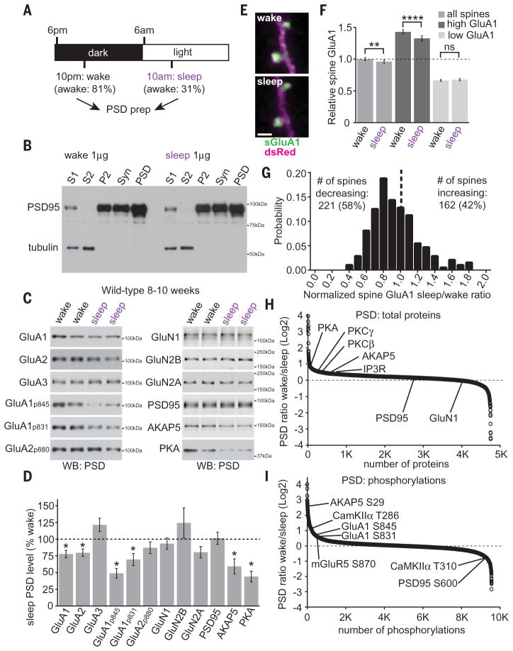
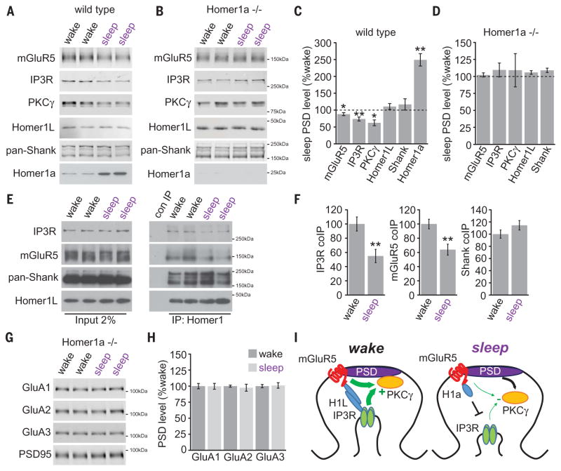
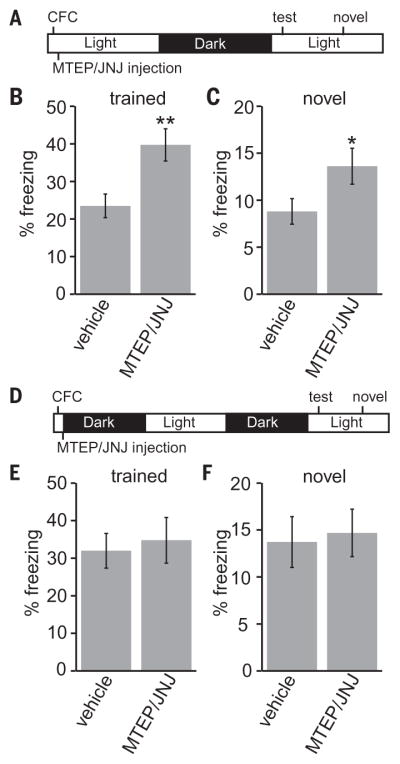
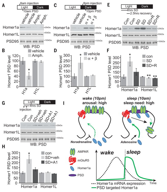
Sleep is an essential process that supports learning and memory by acting on synapses through poorly understood molecular mechanisms. Using biochemistry, proteomics, and imaging in mice, we find that during sleep, synapses undergo widespread alterations in composition and signaling, including weakening of synapses through removal and dephosphorylation of synaptic AMPA-type glutamate receptors. These changes are driven by the immediate early gene Homer1a and signaling from group I metabotropic glutamate receptors mGluR1/5. Homer1a serves as a molecular integrator of arousal and sleep need via the wake- and sleep-promoting neuromodulators, noradrenaline and adenosine, respectively. Our data suggest that homeostatic scaling-down, a global form of synaptic plasticity, is active during sleep to remodel synapses and participates in the consolidation of contextual memory.


Figures

Fig. 1
Fig. 1. Forebrain postsynaptic densities are remodeled during the wake/sleep cycle
(A) Experimental design. Time spent awake was measured during the first 4 hours of the dark or light phase by video recording as a fraction of total time. (B) Subcellular fractionation to yield S1, S2, P2, synaptosome (Syn), and postsynaptic density (PSD) fractions. (C and D) Western blot analysis of wake/sleep PSD fractions. N = 8 brains for each condition; *P < 0.05, Student’s t test. (E) Wake/sleep phase in vivo two-photon imaging of SEP-tagged GluA1 in dendritic spines (sGluA1, green) and dsRed (magenta) in layer V pyramidal neurons of the primary motor cortex. Scale bar, 2 μm. (F) Relative spine GluA1 is significantly reduced during sleep compared with wake. Spines that contain greater-than-average spine GluA1 during wake (high GluA1) show a greater change than spines with lower-than-average spine GluA1 (low GluA1). N = 383 spines from 4 mice; “high GluA1” N = 168 spines; “low GluA1” N = 215 spines; **P < 0.01; ****P < 0.0001; ns, not significant, Student’s t test. (G) Change in spine GluA1 during sleep (sleep/wake ratio) histogram. More spines show a decrease in spine GluA1 during sleep than an increase. N = 383 spines from 4 mice, significantly different from a randomized distribution, Kolmogorov-Smirnov test, P < 0.05. (H and I) Quantitative proteomics of PSD total proteome (H) or phosphoproteome (I). Positive and negative values indicate PSD enrichment during wake or sleep, respectively. Data obtained from 5 mice per condition. Error bars, mean ± SEM.
Fig. 2
Fig. 2. Wake/sleep remodeling of the mGluR5 signaling complex by Homer1a
(A to D) Western blot analysis of mGluR5 signaling proteins in PSD fractions collected from wild-type or Homer1a KO mice during wake or sleep. N = 8 brains for each condition; *P < 0.05, **P < 0.01, Student’s t test. (E and F) Coimmunoprecipitation of mGluR5, IP3R, Shank, and Homer1L in forebrain P2 fractions during wake or sleep. N = 8; **P < 0.01, Student’s t test. (G and H) Western blot analysis of AMPARs in PSD fractions from Homer1a KO mice during wake or sleep. N = 8 brains for each condition; **P < 0.01, Student’s t test. (I) Model of Homer1a-dependent remodeling of mGluR5 signaling complex. Error bars, mean ± SEM.
Fig. 3
Fig. 3. mGluR1/5 signaling during the sleep phase affects memory consolidation
(A to C) Mice were trained in contextual fear conditioning at the beginning of the sleep phase, followed by intraperitoneal injection of vehicle or MJ. Mice were tested 24 hours later for freezing responses in the trained context (B) or a novel context (C). N = 19 vehicle, 19 MJ; *P < 0.05, **P < 0.01, Student’s t test. (D to F) Mice were trained and injected, as above, before the wake phase. Mice were tested for freezing responses >36 hours later at the same time of day as above in the trained context (E) or a novel context (F). N = 12 vehicle, 12 MJ; P > 0.05, Student’s t test. Error bars, mean ± SEM.
Fig. 4
Fig. 4. Homer1a PSD targeting is controlled by NA and adenosine signaling
(A and B) Mice were injected with D-amphetamine (Amph.), 2 mg per kg of weight (mg/kg) at 8 a.m., and forebrain PSD was prepared 2 hours later at 10 a.m. Amphetamine treatment resulted in a significant decrease in Homer1a PSD targeting. N = 5; *P < 0.05, Student’s t test. (C and D) Mice were injected with a cocktail of α and β adrenergic receptor inhibitors, prazosin and propranolol (α + β) (2 mg/kg and 20 mg/kg) at 8 p.m., and forebrain PSD was prepared 2 hours later at 10 p.m. α + β treatment resulted in a large increase in Homer1a PSD targeting. N = 4; *P < 0.05, Student’s t test. (E and F) Mice were sleep deprived (SD) for 4 hours (6 to 10 a.m.) by placing mice into a new cage with or without recovery sleep (2.5 hours, SD+R) in their original cage, followed by forebrain PSD preparation. SD resulted in a significant increase in Homer1a PSD targeting that was reversed by recovery sleep. N = 8; *P < 0.05, **P < 0.01, Student’s t test with Bonferroni correction. (G and H) Mice were left in their home cage (con) or subjected to 4 hours SD (6 to 10 a.m.), with two injections, spaced 2 hours apart, of vehicle or adenosine A1 receptor inhibitor DPCPX (A1) (0.5 mg/kg), followed by PSD isolation and Western blot. A1 blockade prevented up-regulation of Homer1a in the PSD during SD. N = 6; *P < 0.05, Student’s t test with Bonferroni correction. (I) Model. Homer1a targeting to the PSD is inhibited by noradrenaline and promoted by adenosine. Homer1a in the PSD binds to mGluR5 and activates signaling to promote AMPAR removal. Black arrows indicate signaling pathways; green arrows indicate protein trafficking/translocation. Homer1a mRNA is expressed during wake after synaptic plasticity such as LTP and learning. Homer1a mRNA expression is low during sleep. The amount of scaling-down during sleep will be a function of the amount of Homer1a expressed during wake. Error bars, mean ± SEM.

Comment in

  • Synaptic scaling in sleep.
    Acsády L, Harris KD. Acsády L, et al. Science. 2017 Feb 3;355(6324):457. doi: 10.1126/science.aam7917. Science. 2017. PMID: 28154034 No abstract available.

References

    1. Diekelmann S, Born J. Nat Rev Neurosci. 2010;11:114–126. - PubMed
    1. Vecsey CG, et al. Nature. 2009;461:1122–1125. - PMC - PubMed
    1. Vyazovskiy VV, Cirelli C, Pfister-Genskow M, Faraguna U, Tononi G. Nat Neurosci. 2008;11:200–208. - PubMed
    1. Liu ZW, Faraguna U, Cirelli C, Tononi G, Gao XB. J Neurosci. 2010;30:8671–8675. - PMC - PubMed
    1. Tononi G, Cirelli C. Neuron. 2014;81:12–34. - PMC - PubMed

Publication types

MeSH terms