. 2017 Feb 3;355(6324):528-531.
doi: 10.1126/science.aal2415.

The structure of the yeast mitochondrial ribosome

Affiliations

The structure of the yeast mitochondrial ribosome

Nirupa Desai et al. Science. .

Abstract

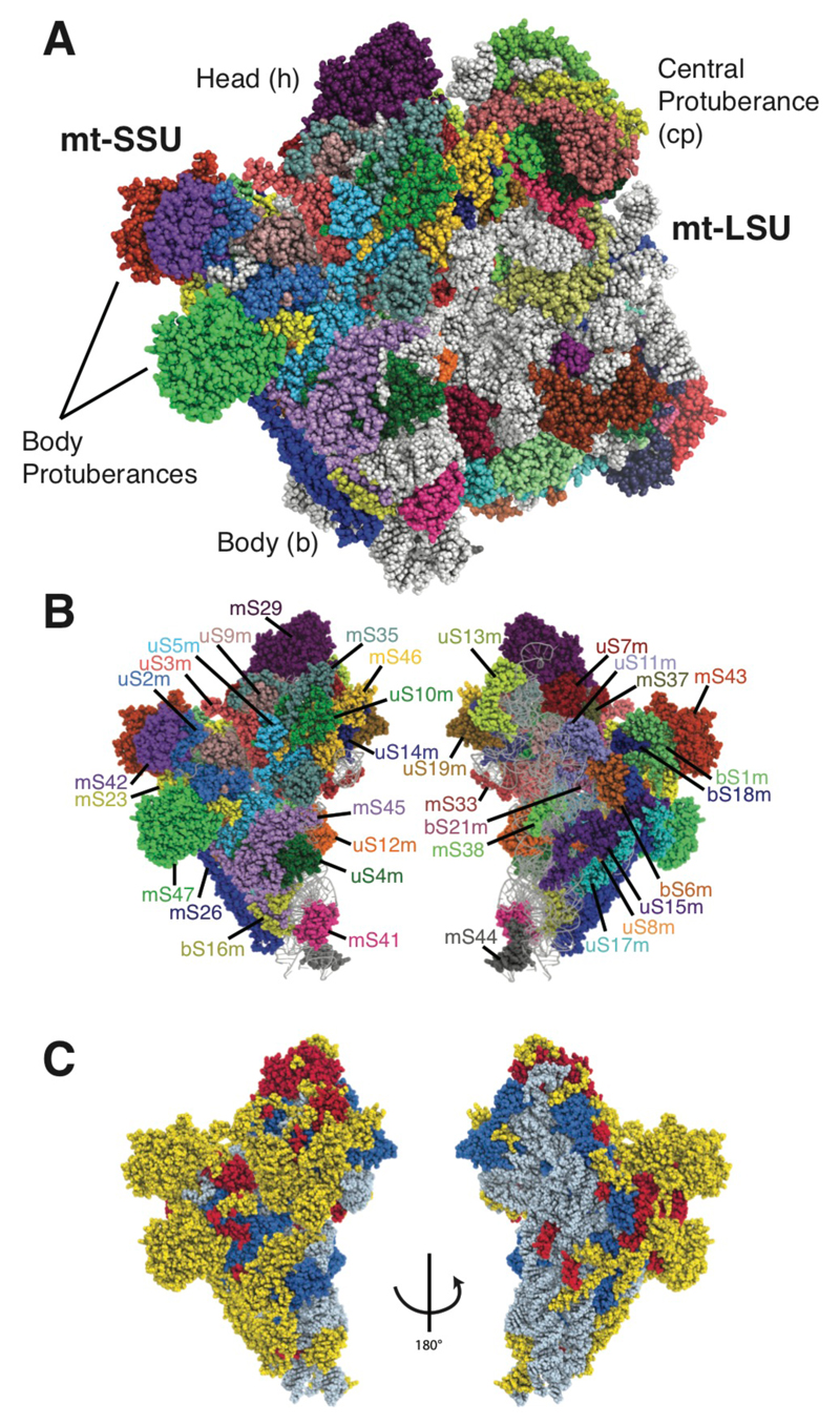
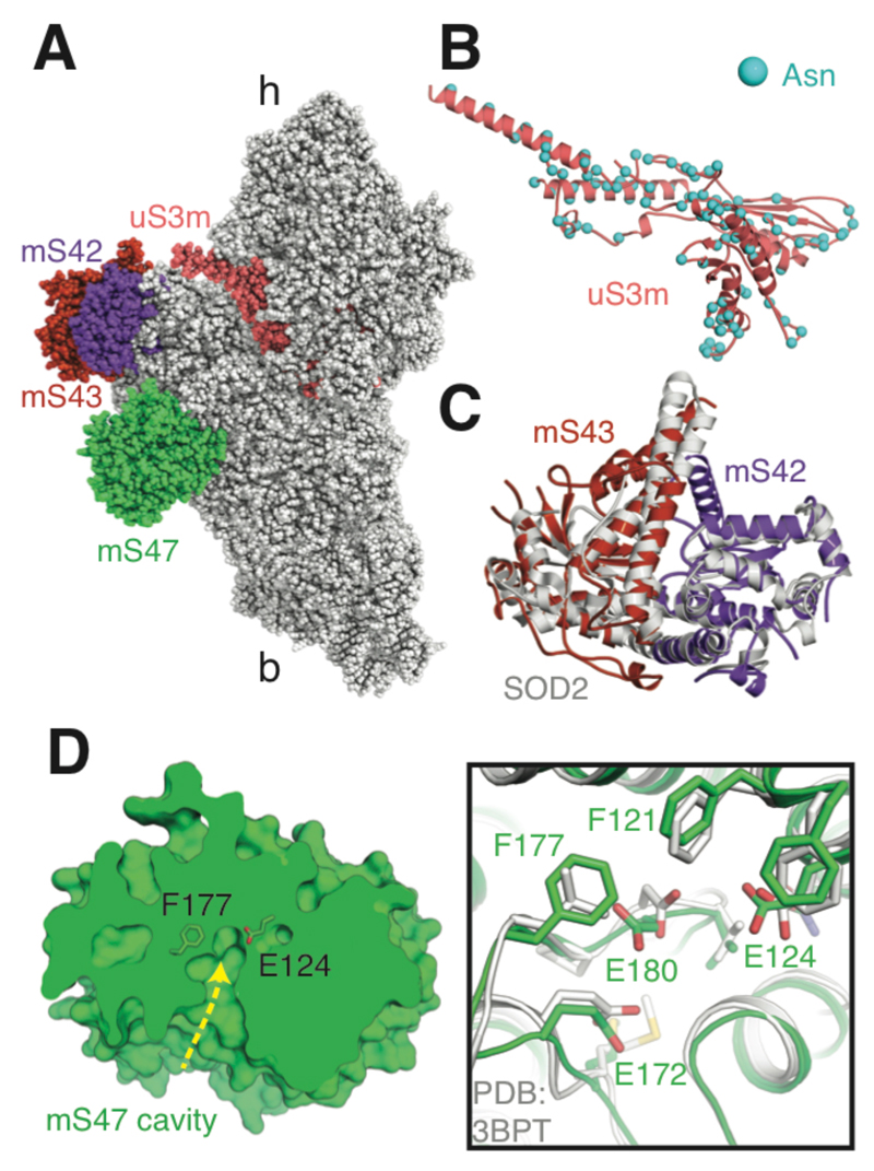
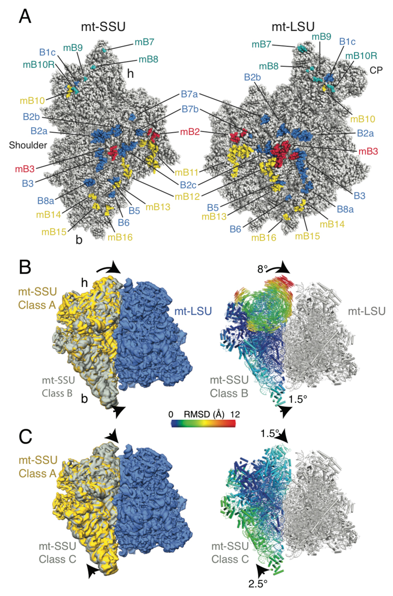
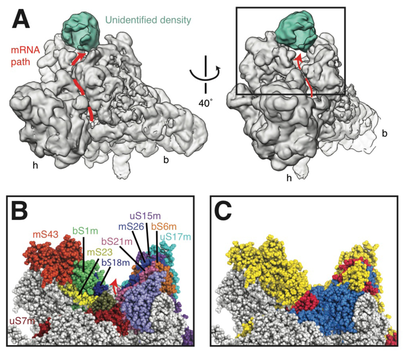
Mitochondria have specialized ribosomes (mitoribosomes) dedicated to the expression of the genetic information encoded by their genomes. Here, using electron cryomicroscopy, we have determined the structure of the 75-component yeast mitoribosome to an overall resolution of 3.3 angstroms. The mitoribosomal small subunit has been built de novo and includes 15S ribosomal RNA (rRNA) and 34 proteins, including 14 without homologs in the evolutionarily related bacterial ribosome. Yeast-specific rRNA and protein elements, including the acquisition of a putatively active enzyme, give the mitoribosome a distinct architecture compared to the mammalian mitoribosome. At an expanded messenger RNA channel exit, there is a binding platform for translational activators that regulate translation in yeast but not mammalian mitochondria. The structure provides insights into the evolution and species-specific specialization of mitochondrial translation.


Figures

Fig. 1
Fig. 1. The structure of the yeast mitoribosome.
(A) The overall structure of the complete yeast mitoribosome. (B) The mitoribosomal proteins of the yeast mt-SSU. (C) Protein elements of the mt-SSU colored by conservation; elements conserved with the bacterial ribosome are blue, elements conserved with the human mitoribosome are red, and elements specific to the yeast mitoribosome are yellow. rRNA is colored grey.
Fig. 2
Fig. 2. Features of the yeast mt-SSU.
(A) The structure of the yeast mt-SSU with uS3m, mS42, mS43 and mS47 highlighted. (B) The asparagine residues of mitochondria-encoded uS3m are distributed on the protein surface. (C) mS42 and mS43 (related to panel A by a 90° rotation around the y axis) form a heterodimer that structurally resembles a yeast mitochondrial superoxide dismutase dimer (Protein Data Bank ID 3LSU). (D) mS47 (related to panel A by an approximate 180° rotation around the x axis) is a probable enzyme with a large cavity and catalytic residues conserved with human β-hydroxyisobutyryl-CoA hydrolase (Protein Data Bank ID 3BPT). Single-letter abbreviations for the amino acid residues are as follows: E, Glu; and F, Phe.
Fig. 3
Fig. 3. Bridges and mitoribosomal dynamics.
(A) Intersubunit interfaces with residues that contribute to bridges highlighted. Bridges also present in the bacterial ribosome are in blue; mitoribosome-specific bridges conserved in the human mitoribosome are red and yeast-specific bridges are in yellow. Residues that form additional bridges in class B are shown in teal. (B) Class B is related to class A by a small rotation of the body and an 8° rotation of the head. The body atoms and head vectors (right) are colored by root-mean-square displacement from their positions in class A. (C) Class C is related to class A by small rotations of the body and head.
Fig. 4
Fig. 4. The extended mRNA channel exit.
(A) View from the mt-LSU, showing the path of the mRNA channel (red line with arrow) around the neck of the yeast mt-SSU. Additional density is located above a canyon at the mRNA channel exit. The boxed section is shown in panels B-C. (B) The proteins that form the canyon. (C) The canyon walls are formed predominantly by mitoribosome-specific protein elements (colored red and yellow as in Fig. 1C).

References

    1. Gray MW, Burger G, Lang BF. Mitochondrial evolution. Science. 1999;283:1476–1481. - PubMed
    1. Ott M, Amunts A, Brown A. Organization and Regulation of Mitochondrial Protein Synthesis. Annu Rev Biochem. 2016;85:77–101. - PubMed
    1. Greber BJ, Ban N. Structure and Function of the Mitochondrial Ribosome. Annu Rev Biochem. 2016;85:103–132. - PubMed
    1. Amunts A, et al. Structure of the yeast mitochondrial large ribosomal subunit. Science. 2014;343:1485–1489. - PMC - PubMed
    1. Amunts A, Brown A, Toots J, Scheres SHW, Ramakrishnan V. Ribosome. The structure of the human mitochondrial ribosome. Science. 2015;348:95–98. - PMC - PubMed

Publication types

MeSH terms