Published Erratum
. 2017 Feb;173(2):1522-1523.
doi: 10.1104/pp.16.01911.

CORRECTION: Vol. 150: 1260-1271, 2009

No authors listed
Published Erratum

CORRECTION: Vol. 150: 1260-1271, 2009

No authors listed. Plant Physiol. 2017 Feb.
No abstract available


Figures

Figure 6.
Figure 6.
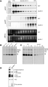
Original: Polysome association and in vivo labeling of wild-type (WT) and lpa66-1 plants. A, Association of psbA and psbD mRNAs with polysomes. Ten fractions of equal volume were collected from the top to bottom of 15% to 55% Suc gradients, and equal proportions of the RNA purified from each fraction were analyzed by gel-blot hybridization. rRNAs were detected by ethidium bromide staining. The RNA size markers are indicated to the left. B, Pulse labeling of thylakoid membrane proteins. After pulse labeling young Arabidopsis seedlings in the presence of cycloheximide for 20 min, thylakoid membranes were isolated, and the proteins were separated by SDS-urea-PAGE and visualized autoradiographically. C, Pulse and chase labeling of thylakoid membrane proteins. After pulse labeling for 20 min followed by 1-, 2-, or 4-h chases with cold Met, thylakoid membranes were isolated, and the proteins were separated by SDS-urea-PAGE and visualized autoradiographically. D, BN gel analysis of labeled thylakoid membrane protein complexes after pulse labeling. After a 20-min pulse in Arabidopsis young seedlings in the presence of cycloheximide, the thylakoid membranes were isolated and solubilized with dodecyl-β-d-maltoside, then the protein complexes were separated by BN-PAGE and visualized autoradiographically. Bands corresponding to various PSII assembly complexes are indicated to the right.
Figure 6.
Figure 6.
Corrected: Polysome association and in vivo labeling of wild-type (WT) and lpa66-1 plants. A, Association of psbA and psbD mRNAs with polysomes. Ten fractions of equal volume were collected from the top to bottom of 15% to 55% Suc gradients, and equal proportions of the RNA purified from each fraction were analyzed by gel-blot hybridization. rRNAs were detected by ethidium bromide staining. The RNA size markers are indicated to the left. The images of the psbD probed blot and its corresponding ethidium bromide-stained gel were from an independent duplicated experiment. B, Pulse labeling of thylakoid membrane proteins. After pulse labeling young Arabidopsis seedlings in the presence of cycloheximide for 20 min, thylakoid membranes were isolated, and the proteins were separated by SDS-urea-PAGE and visualized autoradiographically. C, Pulse and chase labeling of thylakoid membrane proteins. After pulse labeling for 20 min followed by 1-, 2-, or 4-h chases with cold Met, thylakoid membranes were isolated, and the proteins were separated by SDS-urea-PAGE and visualized autoradiographically. D, BN gel analysis of labeled thylakoid membrane protein complexes after pulse labeling. After a 20-min pulse in Arabidopsis young seedlings in the presence of cycloheximide, the thylakoid membranes were isolated and solubilized with dodecyl-β-d-maltoside, then the protein complexes were separated by BN-PAGE and visualized autoradiographically. Bands corresponding to various PSII assembly complexes are indicated to the right.

Erratum for

Publication types

LinkOut - more resources