. 2017 Apr;38(4):721-725.
doi: 10.3174/ajnr.A5085. Epub 2017 Feb 2.

Benign Enhancing Foramen Magnum Lesions: Clinical Report of a Newly Recognized Entity

Affiliations

Benign Enhancing Foramen Magnum Lesions: Clinical Report of a Newly Recognized Entity

B J McGuinness et al. AJNR Am J Neuroradiol. 2017 Apr.

Abstract

Intradural extramedullary foramen magnum enhancing lesions may be due to meningioma, nerve sheath tumor, aneurysm, or meningeal disease. In this clinical report of 14 patients, we describe a novel imaging finding within the foramen magnum that simulates disease. The lesion is hyperintense on 3D-FLAIR and enhances on 3D gradient-echo sequences but is not seen on 2D-TSE T2WI. It occurs at a characteristic location related to the posterior aspect of the intradural vertebral artery just distal to the dural penetration. Stability of this lesion was demonstrated in those patients who underwent follow-up imaging. Recognition of this apparently benign lesion may prevent unnecessary patient anxiety and repeat imaging.


Figures

Fig 1.
Fig 1.
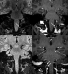
Axial (upper left) and sagittal (upper right) reformats of a 3D-FLAIR sequence show a 6-mm hyperintense lesion within the left posterior aspect of the foramen magnum. This lesion shows enhancement on the axial (lower left) and sagittal (lower right) reformats of the 3D-T1-weighted postcontrast sequences. The lesion lies posteromedial to the dural penetration of the left vertebral artery.
Fig 2.
Fig 2.
Four different examples of foramen magnum lesions. 3D-FLAIR images (upper row) show lesions as hyperintense foci near the posterior aspect of the dural penetration of the vertebral artery (white arrows). Corresponding 3D-T1-weighted postcontrast images (center row) show that the lesions enhance (white arrows). TOF-MRA axial source images (lower row) demonstrate the relationship of local arteries to the lesions, which do not show flow-related signal return.
Fig 3.
Fig 3.
Axial reformats of a 3D-FLAIR (upper left image) sequence show a 4-mm hyperintense focus within the right side of the foramen magnum just posterior to the dural penetration of the right vertebral artery (white arrow). Axial reformat of a 3D-T1-weighted postcontrast sequence (upper center image) shows that this lesion enhances (white arrow). The lesion is isointense with CSF on a TSE T2-weighted (lower left image, black arrow) and hypointense on a TSE T2-weighted image with a 90° flip-back pulse (T2 driven equilibrium radiofrequency reset pulse, lower center image, black arrowhead). The lesion is occult on TOF-MRA (upper right image) and B1000 (lower right image).
Fig 4.
Fig 4.
Two examples of a possible venous vascular connection with the lesion. Coronal FLAIR images (upper and lower left images) show typical hyperintense foci (white arrows). Corresponding coronal reformats of 3D-T1-weighted postcontrast images (upper and lower right images) show small enhancing linear channels (white arrows) connecting with the lesions.
Fig 5.
Fig 5.
Diagram showing the posterior view of the left side of the intradural spinal canal at and just below the level of foramen magnum. The typical location of the foramen magnum lesion is shown by the shaded circle. Relationships of the vertebral artery (VA), dorsal root of the C1 nerve (C1), spinal accessory nerve (Sp A), dentate ligament (DL), posterior spinal artery (asterisk), and spinal cord (C) are shown. Note the typically present communicating branch (arrow) between the spinal accessory nerve and the C1 nerve.

Comment in

References

    1. Samii M, Gerganov VM. Surgery of extra-axial tumor of the cerebral base. Neurosurgery 2008;62:1153–66; discussion 1166–68 - PubMed
    1. Talacchi A, Biroli A, Soda C, et al. . Surgical management of ventral and ventrolateral foramen magnum meningiomas: report on a 64-case series and review of the literature. Neurosurg Rev 2012;35:359–67; discussion 367–68 10.1007/s10143-012-0381-6 - DOI - PubMed
    1. Krings T, Geibprasert S. Spinal dural arteriovenous fistulas. AJNR Am J Neuroradiol 2009;30:639–48 10.3174/ajnr.A1485 - DOI - PMC - PubMed
    1. Rhoton AL. The posterior fossa veins. Neurosurgery 2000;47:S69–92 10.1097/00006123-200009001-00012 - DOI - PubMed
    1. Hetts SW, English JD, Stiver SI, et al. . Bilateral cervical spinal arteriovenous fistulas with intracranial venous drainage mimicking a foramen magnum dural arteriovenous fistula. Interv Neuroradiol 2013;19:483–88 - PMC - PubMed

LinkOut - more resources