Diagnostic Quality of 3D T2-SPACE Compared with T2-FSE in the Evaluation of Cervical Spine MRI Anatomy
- PMID: 28154126
- PMCID: PMC7960240
- DOI: 10.3174/ajnr.A5080
Diagnostic Quality of 3D T2-SPACE Compared with T2-FSE in the Evaluation of Cervical Spine MRI Anatomy
Abstract
Background and purpose: Spinal anatomy has been variably investigated using 3D MRI. We aimed to compare the diagnostic quality of T2 sampling perfection with application-optimized contrasts by using flip angle evolution (SPACE) with T2-FSE sequences for visualization of cervical spine anatomy. We predicted that T2-SPACE will be equivalent or superior to T2-FSE for visibility of anatomic structures.
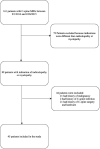
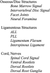
Materials and methods: Adult patients undergoing cervical spine MR imaging with both T2-SPACE and T2-FSE sequences for radiculopathy or myelopathy between September 2014 and February 2015 were included. Two blinded subspecialty-trained radiologists independently assessed the visibility of 12 anatomic structures by using a 5-point scale and assessed CSF pulsation artifact by using a 4-point scale. Sagittal images and 6 axial levels from C2-T1 on T2-FSE were reviewed; 2 weeks later and after randomization, T2-SPACE was evaluated. Diagnostic quality for each structure and CSF pulsation artifact visibility on both sequences were compared by using a paired t test. Interobserver agreement was calculated (κ).
Results: Forty-five patients were included (mean age, 57 years; 40% male). The average visibility scores for intervertebral disc signal, neural foramina, ligamentum flavum, ventral rootlets, and dorsal rootlets were higher for T2-SPACE compared with T2-FSE for both reviewers (P < .001). Average scores for remaining structures were either not statistically different or the superiority of one sequence was discordant between reviewers. T2-SPACE showed less degree of CSF flow artifact (P < .001). Interobserver variability ranged between -0.02-0.20 for T2-SPACE and -0.02-0.30 for T2-FSE (slight to fair agreement).
Conclusions: T2-SPACE may be equivalent or superior to T2-FSE for the evaluation of cervical spine anatomic structures, and T2-SPACE shows a lower degree of CSF pulsation artifact.
© 2017 by American Journal of Neuroradiology.
Figures


Comment in
-
3D T2-SPACE versus T2-FSE or T2 Gradient Recalled-Echo: Which Is the Best Sequence?AJNR Am J Neuroradiol. 2017 Jul;38(7):E48-E49. doi: 10.3174/ajnr.A5190. Epub 2017 Apr 27. AJNR Am J Neuroradiol. 2017. PMID: 28450429 Free PMC article. No abstract available.
-
Reply.AJNR Am J Neuroradiol. 2017 Jul;38(7):E50. doi: 10.3174/ajnr.A5202. Epub 2017 Apr 27. AJNR Am J Neuroradiol. 2017. PMID: 28450430 Free PMC article. No abstract available.
References
-
- Senova S, Hosomi K, Gurruchaga JM, et al. . Three-dimensional SPACE fluid-attenuated inversion recovery at 3 T to improve subthalamic nucleus lead placement for deep brain stimulation in Parkinson's disease: from preclinical to clinical studies. J Neurosurg 2016;125:472–80 10.3171/2015.7.JNS15379 - DOI - PubMed
-
- Czerny C, Trattnig S, Baumgartner WD, et al. . [MRI of the regions of the inner ear and cerebellopontine angle using a 3D T2-weighted turbo spin-echo sequence. Comparison with conventional 2D T2-weighted turbo spin-echo sequences and T1-weighted spin-echo sequences]. [Article in German] Rofo 1997;167:377–83 10.1055/s-2007-1015547 - DOI - PubMed
Publication types
MeSH terms
LinkOut - more resources
Full Text Sources
Other Literature Sources
Medical
Miscellaneous
