. 2017 Feb 14;114(7):1530-1535.
doi: 10.1073/pnas.1621431114. Epub 2017 Feb 1.

pKa of Glu325 in LacY

Affiliations

pKa of Glu325 in LacY

Natalia Grytsyk et al. Proc Natl Acad Sci U S A. .

Abstract

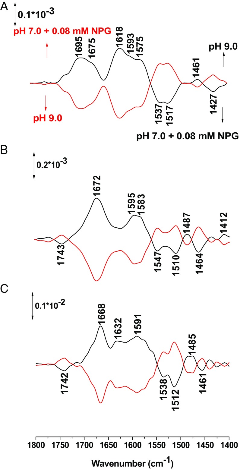
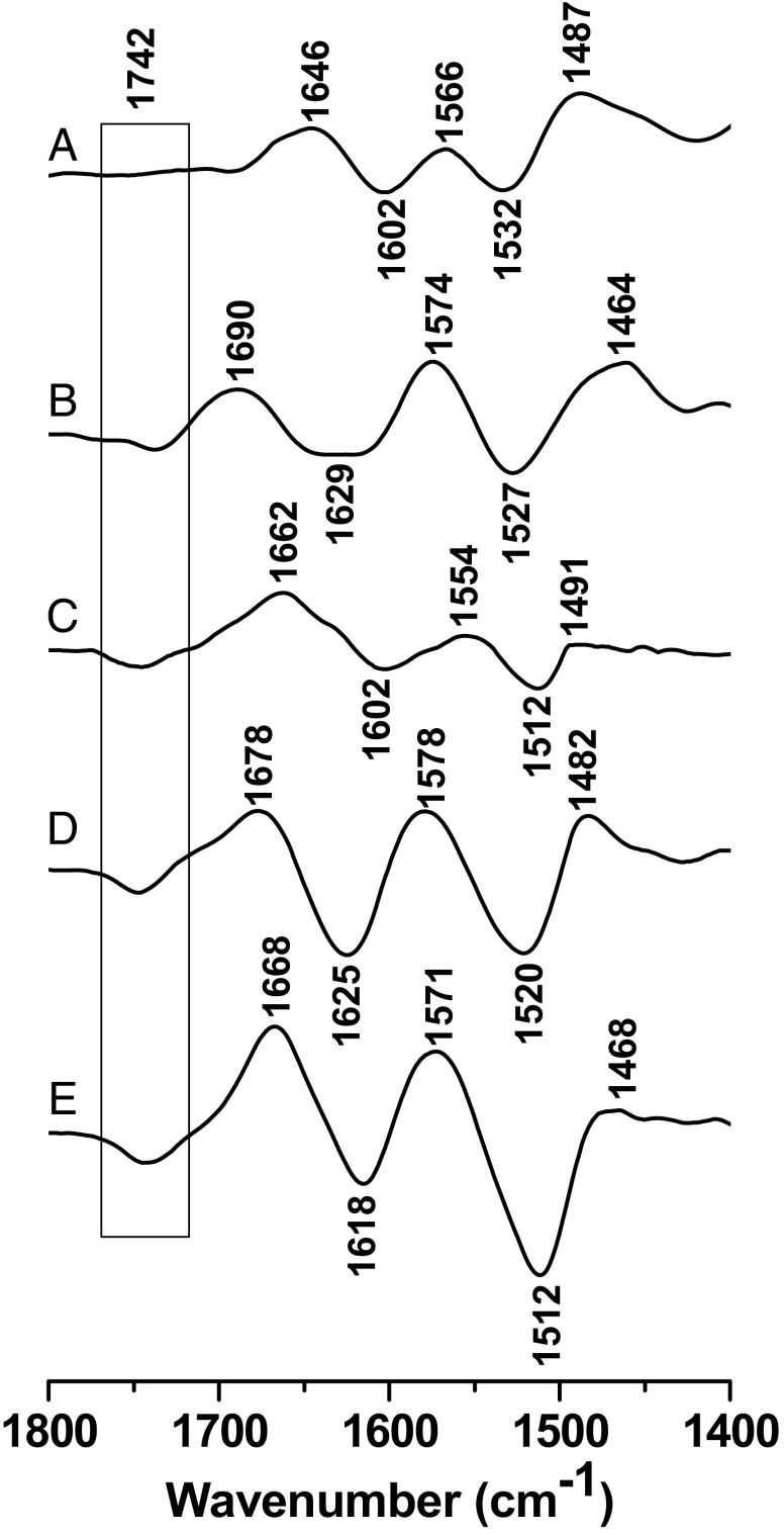
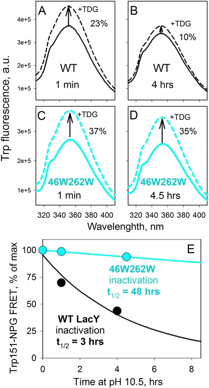
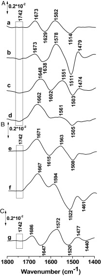
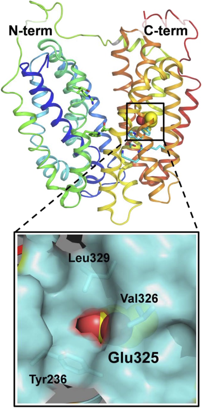
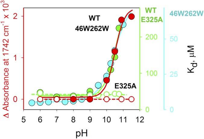
Lactose permease (LacY), a paradigm for the largest family of membrane transport proteins, catalyzes the coupled translocation of a galactoside and a H+ across the cytoplasmic membrane of Escherichia coli (galactoside/H+ symport). One of the most important aspects of the mechanism is the relationship between protonation and binding of the cargo galactopyranoside. In this regard, it has been shown that protonation is required for binding. Furthermore when galactoside affinity is measured as a function of pH, an apparent pK (pKapp) of ∼10.5 is obtained. Strikingly, when Glu325, a residue long known to be involved in coupling between H+ and sugar translocation, is replaced with a neutral side chain, the pH effect is abolished, and high-affinity binding is observed until LacY is destabilized at alkaline pH. In this paper, infrared spectroscopy is used to identify Glu325 in situ. Moreover, it is demonstrated that this residue exhibits a pKa of 10.5 ± 0.1 that is insensitive to the presence of galactopyranoside. Thus, it is apparent that protonation of Glu325 specifically is required for effective sugar binding to LacY.

Keywords: lactose permease; membrane proteins; protonation; surface-enhanced infrared spectroscopy; transport.


Conflict of interest statement

The authors declare no conflict of interest.

Figures

Fig. 1.
Fig. 1.
Perfusion-induced FTIR difference spectra of Lac Y obtained from the sample equilibrated at pH 7 in the presence of NPG subtracted from the sample equilibrated at pH 9 (A), 10 (B), and 10.5 (C), respectively (black line) and reverse (red line).
Fig. S1.
Fig. S1.
Immobilization of the protein on the silicon crystal. AD describe the observed reactions and the surface-enhanced infrared spectra measured in order to follow the reaction.
Fig. S2.
Fig. S2.
Absorption IR spectra of LacY and E325 mutant.
Fig. 2.
Fig. 2.
Perfusion-induced FTIR difference spectra of LacY wild type equilibrated in the presence of 0.08 mM NPG at pH 7 subtracted from the sample equilibrated at pH 9.0 (A), 10.0 (B), and 10.5 (C), respectively.
Fig. 3.
Fig. 3.
Perfusion-induced FTIR difference spectra of LacY G46W/G262W in the presence of NPG in both perfusion solutions. The sample equilibrated at pH 7.0 was subtracted from the sample equilibrated at 9.0 (A), 10.0 (B), 10.5 (C), 10.9 (D), and 11.5 (E), respectively.
Fig. S3.
Fig. S3.
Stability of WT LacY and 46W/262W mutant at pH 10.5. Sugar binding was measured as Trp151-NPG Förster resonance energy transfer (44) after preincubation of reconstituted PLs (with 14 mM protein at a lipid-to-protein ratio of 5 (wt/wt) in 50 mM CAPS pH 10.5 for given time at room temperature. (A–D) Increase of Trp fluorescence intensity (excitation at 295 nm) resulting from displacement of bound NPG (0.1 mM) by excess of TDG (6 mM) measured at pH 7.5 in 50 mM NaPi with 0.4 mM LacY. (E) Time course of LacY inactivation at pH 10.5.
Fig. 4.
Fig. 4.
Perfusion-induced FTIR difference spectra of LacY G46W/G262W in the absence of NPG obtained from samples equilibrated at pH 7 subtracted from the samples equilibrated at pH 8.0 (A,a), 10.5 (A, b), 10.9 (A, c), and 11.5 (A, d), respectively. Difference spectra of the LacY G46W/G262W/E325A variant for the step from to pH 7.0 to pH 11.5 (B, e) and 10.9 (B, f), and for the simple LacY E325A mutant for the step from pH 6.0 to pH 10.0 (C, g).
Fig. 5.
Fig. 5.
Position of Glu325 in C-terminal six-helix bundle of WT LacY (Protein Data Bank ID code 2V8N). LacY is presented as rainbow-colored backbone (from blue to red for helices 1–12) with hydrophilic cavity open to cytoplasmic side. Side chain of Glu325 located in helix X is shown as spheres. The area around Glu325 is enlarged with hydrophobic environment displayed as a space-filled cartoon (cyan).
Fig. 6.
Fig. 6.
pH dependence of Δ-IR intensity change at 1,742 cm−1 measured with LacYww in the absence or presence of NPG (filled red circles) or E325A LacY (open red circles), and Kd values for NPG binding to WT LacY (filled green circles), E325A LacY (open green circles), or LacYww (filled cyan circles). Kd values were calculated as the ratio of rate constants (koff/kon) measured by stopped flow (12, 35).

References

    1. Saier MH, Jr, et al. The major facilitator superfamily. J Mol Microbiol Biotechnol. 1999;1(2):257–279. - PubMed
    1. Kaback HR. A chemiosmotic mechanism of symport. Proc Natl Acad Sci USA. 2015;112(5):1259–1264. - PMC - PubMed
    1. Abramson J, et al. Structure and mechanism of the lactose permease of Escherichia coli. Science. 2003;301(5633):610–615. - PubMed
    1. Mirza O, Guan L, Verner G, Iwata S, Kaback HR. Structural evidence for induced fit and a mechanism for sugar/H+ symport in LacY. EMBO J. 2006;25(6):1177–1183. - PMC - PubMed
    1. Guan L, Mirza O, Verner G, Iwata S, Kaback HR. Structural determination of wild-type lactose permease. Proc Natl Acad Sci USA. 2007;104(39):15294–15298. - PMC - PubMed

Publication types

MeSH terms

LinkOut - more resources