. 2017 Mar 17;292(11):4434-4445.
doi: 10.1074/jbc.M116.774109. Epub 2017 Jan 31.

CD44 Transmembrane Receptor and Hyaluronan Regulate Adult Hippocampal Neural Stem Cell Quiescence and Differentiation

Affiliations

CD44 Transmembrane Receptor and Hyaluronan Regulate Adult Hippocampal Neural Stem Cell Quiescence and Differentiation

Weiping Su et al. J Biol Chem. .

Abstract

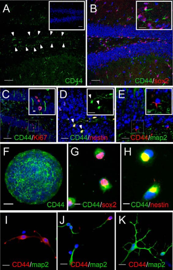
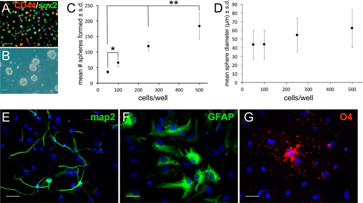
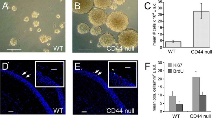
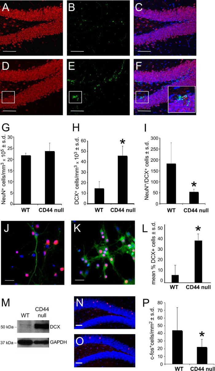
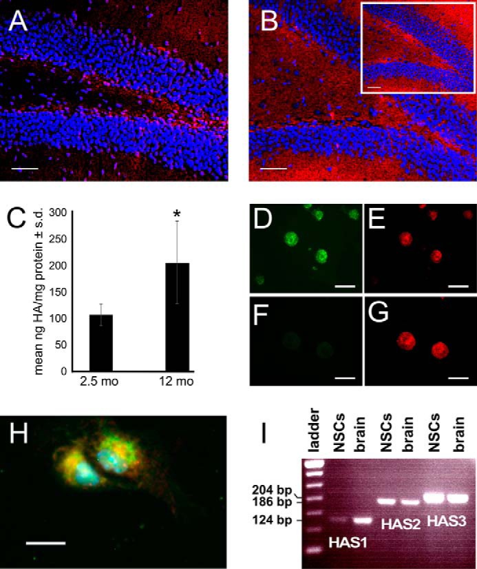
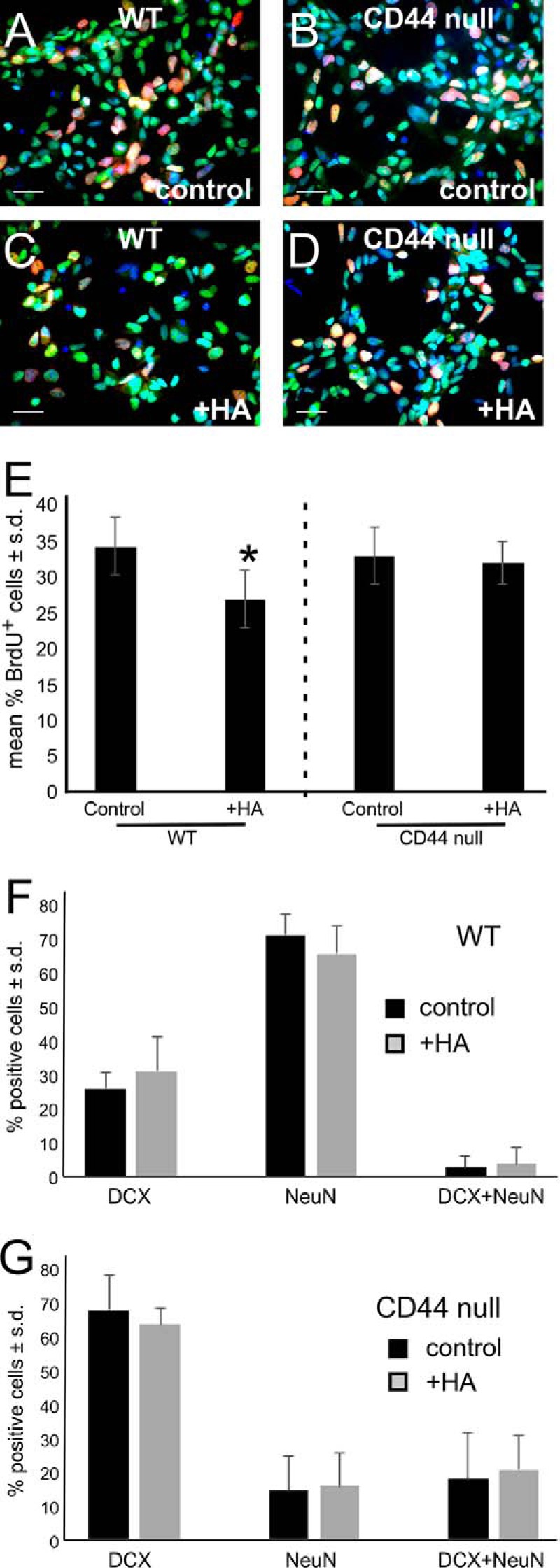
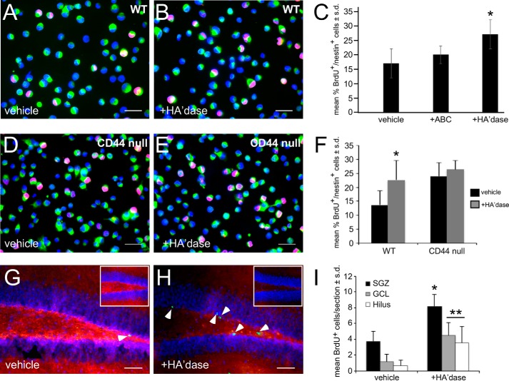
Adult neurogenesis in the hippocampal subgranular zone (SGZ) is involved in learning and memory throughout life but declines with aging. Mice lacking the CD44 transmembrane receptor for the glycosaminoglycan hyaluronan (HA) demonstrate a number of neurological disturbances including hippocampal memory deficits, implicating CD44 in the processes underlying hippocampal memory encoding, storage, or retrieval. Here, we found that HA and CD44 play important roles in regulating adult neurogenesis, and we provide evidence that HA contributes to age-related reductions in neural stem cell (NSC) expansion and differentiation in the hippocampus. CD44-expressing NSCs isolated from the mouse SGZ are self-renewing and capable of differentiating into neurons, astrocytes, and oligodendrocytes. Mice lacking CD44 demonstrate increases in NSC proliferation in the SGZ. This increased proliferation is also observed in NSCs grown in vitro, suggesting that CD44 functions to regulate NSC proliferation in a cell-autonomous manner. HA is synthesized by NSCs and increases in the SGZ with aging. Treating wild type but not CD44-null NSCs with HA inhibits NSC proliferation. HA digestion in wild type NSC cultures or in the SGZ induces increased NSC proliferation, and CD44-null as well as HA-disrupted wild type NSCs demonstrate delayed neuronal differentiation. HA therefore signals through CD44 to regulate NSC quiescence and differentiation, and HA accumulation in the SGZ may contribute to reductions in neurogenesis that are linked to age-related decline in spatial memory.

Keywords: CD44; hippocampus; hyaluronan; neural stem cell (NSC); neurogenesis.


Conflict of interest statement

The authors declare that they have no conflicts of interest with the contents of this article

Figures

FIGURE 1.
FIGURE 1.
CD44 is expressed by NSCs in the SGZ. A, CD44 (green) is expressed by cells in the SGZ (arrowheads). The inset shows CD44 staining in a section of the dentate gyrus from a CD44-null mouse. B, CD44 (green) is expressed by SOX2 (red)-immunolabeled cells. The inset is a magnified image of the area outlined in the left portion of the figure. C, a section showing CD44+ processes (green) of cells with Ki67+ nuclei (red) in both the SGZ and granule cell layer. The area in the inset is an enlargement of the boxed area in the lower portion of the figure. D, a section demonstrating cell bodies that express both nestin (red) and CD44 (green). The inset shows the CD44 channel alone. E, a section showing a lack of CD44 (red) and MAP2 (green) co-localization, indicating that CD44 is not expressed by the granule cells derived from CD44+ NSCs. The inset is an enlargement of the boxed area in the lower left portion of the figure. F, whole mount immunolabeling for CD44 (green) of a neurosphere grown in vitro. G, acutely dissociated NSCs from neurosphere cultures immunolabeled for CD44 (green) and SOX2 (red). H, acutely dissociated NSCs from neurosphere cultures immunolabeled for CD44 (green) and nestin (red). The yellow signal indicates areas where CD44 and nestin co-localize. In all of the images, cells and tissues were stained with Hoechst 33342 (blue; to stain cell nuclei). I–K, CD44 expression declines as SGZ NSCs differentiate into neurons. SGZ-derived NSCs were grown in conditions that favored neuronal differentiation for 1 (I), 3 (J), and 6 days (K). Cells were stained with Hoechst 33342 (blue) to stain cell nuclei and antibodies against CD44 (red) and MAP2 (green). Note that as cells differentiate into more mature neurons they lose CD44 expression. Scale bars, 50 (A–C and inset in A), 15 (insets in B and D, D–F, and I–K), and 5 μm (G and H).
FIGURE 2.
FIGURE 2.
CD44-expressing NSCs are self-renewing, multipotent cells. A, image of freshly sorted, CD44+ SGZ-derived NSCs immunolabeled with antibodies against SOX2 (green) and CD44 (red) and stained with Hoechst 33342 (blue) to label cell nuclei. B, secondary neurospheres formed from sorted CD44+ NSCs. C, quantification of sphere forming assay as a function of NSC plating density. *, p < 0.001; **, p < 0.05. D, quantification of sphere diameter as a function of NSC plating density. Note there was no significant difference in mean sphere diameters with increasing plating density. E, MAP2 immunolabeling (green) in a culture of CD44+ NSCs grown in neuron differentiation medium. F, glial fibrillary acidic protein (GFAP) immunolabeling (green) in a culture of CD44+ NSCs grown in astrocyte differentiation medium. G, O4 immunolabeling (red) in a culture of CD44+ NSCs grown in oligodendrocyte differentiation medium. In each experiment (E–G), cells were stained with Hoechst 33342 (blue) to label cell nuclei. Scale bars, 25 (A and E–G) and 100 μm (B). Error bars represent S.D.
FIGURE 3.
FIGURE 3.
NSCs from CD44-null mice hyperproliferate. A, SGZ neurospheres from 2-month-old WT mice grown for 6 days. B, SGZ neurospheres from 2-month-old CD44-null mice plated at the same density as the cells in A and grown for 6 days. C, quantification of total cell numbers in neurosphere cultures from WT and CD44-null mice (n = 4). *, p < 0.001. D, section through 9-month-old WT dentate gyrus following a 3-h BrdU pulse. Sections are immunolabeled with an anti-BrdU antibody (red), an anti-Ki67 antibody (green), and Hoechst 33342 (blue; to label cell nuclei). The inset is an enlarged view of the area indicated by the arrows. E, section through a 9-month-old CD44-null dentate gyrus immunolabeled as described in D. The inset is an enlarged view of the area indicated by the arrows. F, quantification of BrdU+ and Ki67+ cells in sections through the dentate gyrus of 9-month-old WT and CD44-null mice (n = 6). pos., positive. *, p < 0.01; **, p < 0.02. Scale bars, 100 (A and B), 50 (D and E), and 25 μm (insets in D and E). Error bars represent S.D.
FIGURE 4.
FIGURE 4.
Neuronal differentiation of CD44-null NSCs is delayed. A, NeuN staining (red) in the dentate gyrus of a 9-month-old WT mouse. B, DCX staining (green) in the same section shown in A. C, merged NeuN and DCX immunolabeling from A and B. The section was also stained with Hoechst 33342 (blue) to label cell nuclei. D, NeuN (red) immunolabeling in the dentate gyrus of a 9-month-old CD44-null mouse. E, DCX staining (green) in the same section shown in D. F, merged NeuN and DCX immunolabeling from D and E co-stained with Hoechst 33342 (blue). The insets in F are magnified areas outlined in the lower left portion of each figure. G, quantification of NeuN immunolabeling in the dentate gyrus of WT and CD44-null mice (n = 6) by unbiased stereological analysis. H, quantification of DCX immunolabeling in the dentate gyrus of WT and CD44-null mice (n = 6) by unbiased stereological analysis. *, p < 0.01. I, quantification of the ratios of NeuN:DCX immunolabeling in the dentate gyrus of WT and CD44-null mice. *, p < 0.001. J, acutely dissociated WT SGZ neurosphere cultures grown in neuronal differentiation medium and immunolabeled with NeuN (red) and DCX (green). Cells were also stained with Hoechst 33342 (blue) to label cell nuclei. K, acutely dissociated CD44-null SGZ neurosphere cultures grown in neuronal differentiation medium and immunolabeled as in J. L, quantification of DCX:NeuN ratios in WT and CD44-null NSC cultures grown in neuronal differentiation medium. *, p < 0.001. M, Western blot showing higher DCX expression in CD44-null as compared with WT SGZ-derived NSC cultures undergoing neuronal differentiation. Blots were probed for GAPDH as a loading control. N, dentate gyrus section from a 6-month-old WT mouse immunolabeled with an anti-c-Fos antibody (red) following behavioral training. The section was also stained with Hoechst 33342 (blue) to label cell nuclei. O, dentate gyrus section from a 6-month-old CD44-null mouse immunolabeled with an anti-c-Fos antibody as in N following behavioral training. P, quantification of c-Fos immunolabeling in the dentate gyrus of WT and CD44-null mice (n = 5). *, p < 0.001. Scale bars, 50 (A–F, N, and O) and 25 μm (J and K). Error bars represent S.D.
FIGURE 5.
FIGURE 5.
SGZ NSCs synthesize HA. A, section of dentate gyrus from a WT 3-month-old mouse labeled with a biotinylated HA-binding protein (red). The section was also stained with Hoechst 33342 (blue) to label cell nuclei. B, section of dentate gyrus from a WT 9-month-old mouse labeled with a biotinylated HA-binding protein as in A. The inset shows HA staining in a 9-month-old CD44-null mouse, demonstrating that HA accumulates in the dentate gyrus independently of CD44 expression. C, quantification of HA in young (2.5-month-old) and old (12-month-old) mouse dentate gyrus tissue lysates using an ELISA-based assay. *, p < 0.001. D, whole mount labeling for CD44 (green) of a WT neurosphere grown in vitro. E, whole mount labeling for HA (red) of the neurospheres shown in D. F, whole mount labeling for CD44 (green) of a CD44-null neurosphere grown in vitro. Note that only background signal is detectable. G, whole mount labeling for HA (red) in the neurospheres shown in F. H, WT NSCs were acutely dissociated from neurospheres and labeled with an anti-nestin antibody (green) and a biotinylated HA-binding protein (red). Cells were also stained with Hoechst 33342 (blue). I, RT-PCR for HAS1, HAS2, and HAS3 of RNA from WT mouse NSCs and WT 2-month-old mouse whole brain. Scale bars, 50 (A and B), 100 (D–G), and 15 μm (H). Error bars represent S.D.
FIGURE 6.
FIGURE 6.
HA blocks NSC proliferation in a CD44-dependent manner. A–D, BrdU (red) and SOX2 (green) double labeling in wild type (A and C) and CD44-null (B and D) NSCs treated with vehicle (A and B) or 100 μg/ml HA (C and D). E, quantification of the percentage of BrdU+/SOX2+ HA-treated WT versus CD44-null NSCs compared with controls (cultures treated with vehicle). *, p < 0.001. F, quantification of the effects of HA on WT NSC neuronal differentiation (as assessed by DCX and NeuN immunolabeling) compared with vehicle controls. G, quantification of the effects of HA on CD44-null NSC neuronal differentiation compared with vehicle controls. Scale bars, 25 μm (A–D). Experiments were each performed three times. Error bars represent S.D.
FIGURE 7.
FIGURE 7.
Digestion of HA surrounding NSCs induces NSC proliferation. A, WT NSCs were pulsed with BrdU after 3 days in vitro in the presence of vehicle (PBS). Cells were then immunolabeled with an anti-BrdU antibody (red) and an anti-nestin antibody (green). Cells were also stained with Hoechst 33342 (blue). B, WT NSCs were pulsed with BrdU after 3 days in vitro in the presence of hyaluronidase. Cells were immunolabeled for BrdU and nestin as in A. C, quantification of BrdU labeling in control and rPH20 (20 units/ml; HA'dase)- or chondroitinase ABC (0.1 units/ml)-treated SGZ NSC cultures (mean percentage of positive cells). *, p < 0.004. D, CD44-null NSCs were pulsed with BrdU after 3 days in vitro in the presence of vehicle (PBS). Cells were then immunolabeled with an anti-BrdU antibody (red) and an anti-nestin antibody (green). Cells were also stained with Hoechst 33342 (blue). E, CD44-null NSCs were pulsed with BrdU after 3 days in vitro in the presence of rPH20. Cells were immunolabeled for BrdU and nestin as in D. F, quantification of BrdU labeling in WT and CD44-null NSC cultures treated with vehicle or rPH20 (mean percentage of positive cells). *, p < 0.004. G, dentate gyrus section from a 6-month-old WT mouse stained with a biotinylated HA-binding protein (red) and an anti-BrdU antibody (green; arrowhead) 2 days following stereotactic injection of vehicle (PBS) and administration of BrdU. Sections were also stained with Hoechst 33342 (blue). Inset, dentate gyrus section from a 6-month-old WT mouse stained with a biotinylated HA-binding protein (red) 24 h following stereotactic injection of vehicle (PBS). Sections were also stained with Hoechst 33342 (blue). H, dentate gyrus section stained as in G from a 6-month-old WT mouse 2 days following stereotactic injection of rPH20. BrdU labeling was observed in the hilus and SGZ as well as in the inner face of the granule cell layer (arrowheads). Inset, dentate gyrus section stained as in G from a 6-month-old WT mouse 24 h following stereotactic injection of rPH20. I, quantification of BrdU labeling in different regions (hilus, SGZ, and granule cell layer) of dentate gyrus sections from animals treated with vehicle or rPH20 (n = 8). *, p < 0.001; **, p < 0.005. Scale bars, 25 (A, B, D, and E) and 50 μm (G and H). Experiments were each performed three times. Error bars represent S.D.
FIGURE 8.
FIGURE 8.
Hyaluronidase delays neuronal maturation of WT but not CD44-null NSCs. A, acutely dissociated WT SGZ neurosphere cultures grown in neuronal differentiation medium with vehicle and immunolabeled with NeuN (red) and DCX (green). Cells were also stained with Hoechst 33342 (blue) to label cell nuclei. B, acutely dissociated WT SGZ neurosphere cultures grown and immunolabeled as in A but treated with rPH20. C, quantification (mean percentage of positive cells) of DCX+, NeuN+, and double positive immunolabeling in WT NSC cultures undergoing neuronal differentiation and treated with vehicle (PBS) or rPH20. *, p < 0.001; **, p < 0.05. D, quantification of DCX and NeuN immunolabeling as in C in CD44-null NSC cultures undergoing neuronal differentiation and treated with vehicle (PBS) or rPH20. Scale bars, 50 μm (A and B). Error bars represent S.D.

Similar articles

Cited by

References

    1. Spalding K. L., Bergmann O., Alkass K., Bernard S., Salehpour M., Huttner H. B., Boström E., Westerlund I., Vial C., Buchholz B. A., Possnert G., Mash D. C., Druid H., and Frisén J. (2013) Dynamics of hippocampal neurogenesis in adult humans. Cell 153, 1219–1227 - PMC - PubMed
    1. Ming G. L., and Song H. (2011) Adult neurogenesis in the mammalian brain: significant answers and significant questions. Neuron 70, 687–702 - PMC - PubMed
    1. Deng W., Saxe M. D., Gallina I. S., and Gage F. H. (2009) Adult-born hippocampal dentate granule cells undergoing maturation modulate learning and memory in the brain. J. Neurosci. 29, 13532–13542 - PMC - PubMed
    1. Kuhn H. G., Dickinson-Anson H., and Gage F. H. (1996) Neurogenesis in the dentate gyrus of the adult rat: age-related decrease of neuronal progenitor proliferation. J. Neurosci. 16, 2027–2033 - PMC - PubMed
    1. Bondolfi L., Ermini F., Long J. M., Ingram D. K., and Jucker M. (2004) Impact of age and caloric restriction on neurogenesis in the dentate gyrus of C57BL/6 mice. Neurobiol. Aging 25, 333–340 - PubMed

Publication types

LinkOut - more resources