. 2017 Mar 10;292(10):4302-4312.
doi: 10.1074/jbc.M116.773085. Epub 2017 Jan 31.

Transforming Growth Factor-β (TGF-β) Directly Activates the JAK1-STAT3 Axis to Induce Hepatic Fibrosis in Coordination with the SMAD Pathway

Affiliations

Transforming Growth Factor-β (TGF-β) Directly Activates the JAK1-STAT3 Axis to Induce Hepatic Fibrosis in Coordination with the SMAD Pathway

Liu-Ya Tang et al. J Biol Chem. .

Abstract

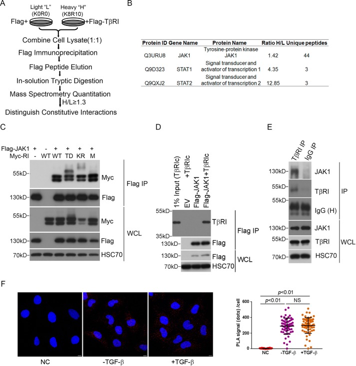
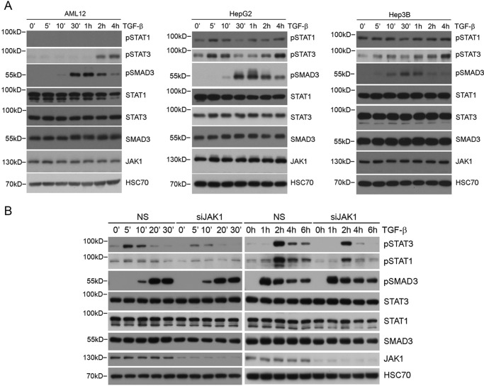
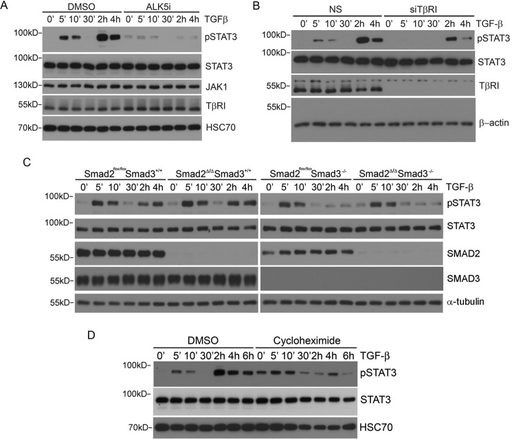
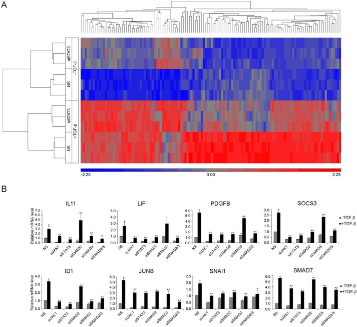
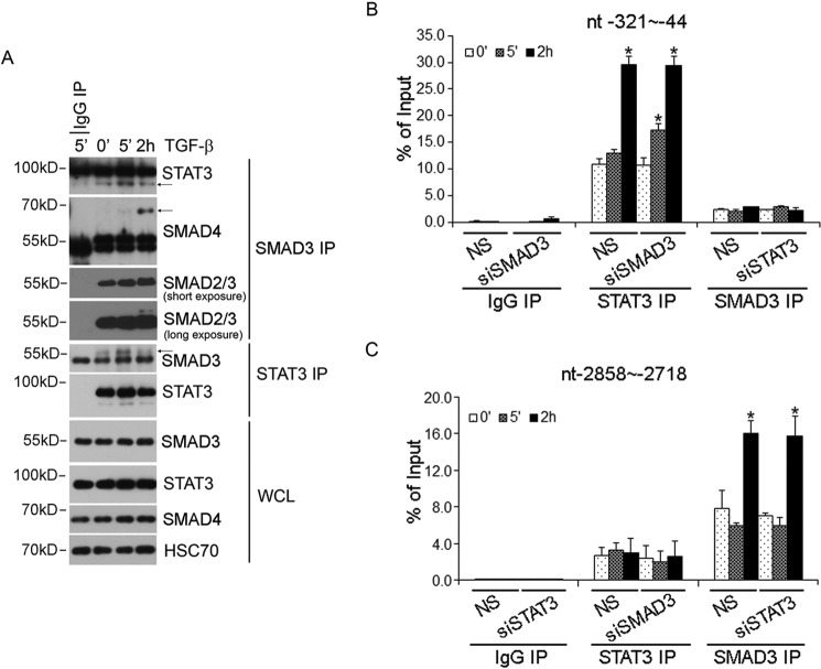
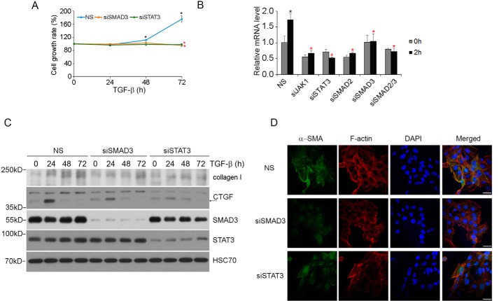
Transforming growth factor-β (TGF-β) signals through both SMAD and non-SMAD pathways to elicit a wide array of biological effects. Existing data have shown the association and coordination between STATs and SMADs in mediating TGF-β functions in hepatic cells, but it is not clear how STATs are activated under these circumstances. Here, we report that JAK1 is a constitutive TGFβRI binding protein and is absolutely required for phosphorylation of STATs in a SMAD-independent manner within minutes of TGF-β stimulation. Following the activation of SMADs, TGF-β also induces a second phase of STAT phosphorylation that requires SMADs, de novo protein synthesis, and contribution from JAK1. Our global gene expression profiling indicates that the non-SMAD JAK1/STAT pathway is essential for the expression of a subset of TGF-β target genes in hepatic stellate cells, and the cooperation between the JAK1-STAT3 and SMAD pathways is critical to the roles of TGF-β in liver fibrosis.

Keywords: Janus kinase (JAK); Liver fibrosis; SMAD transcription factor; STAT3; Smad3; hepatic stellate cell (HSC); transforming growth factor beta (TGF-B).


Conflict of interest statement

The authors declare that they have no conflicts of interest with the contents of this article

Figures

FIGURE 1.
FIGURE 1.
Identification of JAK1 as a TGFβRI-interacting protein by quantitative proteomic analysis. A, SILAC experiment to discover TβRI-interacting proteins. AML12 cells were labeled with l-lysine and l-arginine (K0R0, light) or (K8R10, heavy), respectively. The cells cultured in K8R10 medium (heavy) were transfected with FLAG-TGFβRI, whereas the cells cultured in K0R0 medium (light) were transfected with an empty vector. The cell lysates were combined at equal protein amounts and subjected to MS quantitation. A cut-off of 1.3 heavy/light (H/L) protein ratio was set to weed out background contaminants. B, JAK1, STAT1, and STAT2 were identified as potential TGFβRI-interacting proteins from the screen. C, JAK1 interacts with Myc-tagged wild type TGFβRI as well as TGFβRI mutants in HEK293 cells. TD, constitutively active; KR, kinase-dead; M (mL45), SMAD binding-defective. D, JAK1 interacts with purified cytoplasmic domain of TGFβRI in vitro. E, JAK1 interacts with TGFβRI endogenously. AML12 cells were subjected to immunoprecipitation by TGFβRI antibody or control IgG. The presence of JAK1 in the immunoprecipitated protein complex was detected by Western blotting. F, PLA was used to detect the in vivo interaction of JAK1 and TGFβRI in LX-2 cells. LX-2 cells were treated with ±4 ng/ml TGF-β for 5 min, and the cells were incubated with both JAK1 monoclonal antibody and TGFβRI polyclonal antibody. Negative control (NC) cells were incubated with TGFβRI polyclonal antibody and normal mouse IgG. Scale bar, 10 μm.
FIGURE 2.
FIGURE 2.
TGF-β-induced STAT3 early phosphorylation requires JAK1. A, TGF-β induced STAT3 phosphorylation in different hepatic cells. B, TGF-β induced biphasic phosphorylation of STAT3 in LX-2 cells, and the early phase of STAT3 phosphorylation is dependent on JAK1. LX-2 cells transfected with non-silencing (NS) control or JAK1 siRNA were treated with 4 ng/ml TGF-β for the indicated times.
FIGURE 3.
FIGURE 3.
TGF-β-induced STAT3 early phosphorylation requires TGFβRI but not SMADs. A, kinase activity of TGFβRI is required for TGF-β-induced STAT3 phosphorylation. LX-2 cells were preincubated with 10 μm SB431542 (ALK5i) for 4 h and then treated with 4 ng/ml TGF-β for the indicated times. B, knockdown of TGFβRI in LX-2 cells decreased TGF-β-induced STAT3 phosphorylation. LX-2 cells transfected with the indicated siRNA were treated with 4 ng/ml TGF-β, and cell lysates were subjected to Western blotting. C, the early phase of TGF-β-induced STAT3 phosphorylation is independent of SMAD2/3, but the second phase of activation requires SMAD3. Smad2flox/floxSmad3+/+ or Smad2flox/floxSmad3−/− MEFs were infected with either adeno-GFP or adeno-CRE to generate control or cells devoid of SMAD2 (Smad2Δ/Δ). Two days after adenovirus infection, cells were treated with 4 ng/ml TGF-β for the indicated times, and phospho-STAT3 was detected by Western blotting. D, de novo protein synthesis is required for the TGF-β-induced second phase of STAT3 phosphorylation. LX-2 cells were preincubated with 10 ng/ml cycloheximide (CHX) for 2 h, and the cells were treated with TGF-β for the indicated times.
FIGURE 4.
FIGURE 4.
STAT3 is required for TGF-β-induced transcription of a subset of genes in LX-2 cells. A, hierarchical clustering of TGF-β-responsive and STAT3-dependent genes. Triplicates for each group are shown in the graph. Cells were treated with or without TGF-β for 2 h. B, qRT-PCR results of representative STAT3-dependent TGF-β target genes. Results are shown as relative expression ± S.D. (error bars) (n = 3). Statistically significant difference (p < 0.05) after TGF-β treatment is indicated by a black asterisk; statistically significant differences (p < 0.05) between control and knockdown cells after TGF-β treatment are indicated by a red asterisk.
FIGURE 5.
FIGURE 5.
Interplay between STAT3 and SMAD3 in LX-2 cells. A, physical interaction between STAT3 with SMAD3 in LX-2 cells. LX-2 cells were treated with 4 ng/ml TGF-β for 5 min or 2 h, and SMAD3 or STAT3 protein complex was immunoprecipitated by SMAD3 or STAT3 antibody. B, ChIP analysis of SMAD3 and STAT3 binding to the STAT3 DNA binding sites (bp −321 to −44) in the JUNB promoter after TGF-β stimulation. C, ChIP analysis of SMAD3 and STAT3 binding to the SMAD3 DNA binding sites (bp −2858 to −2718) in the JUNB promoter after TGF-β stimulation. Rabbit IgG was used as a negative control. *, statistically significant differences (p < 0.05) upon TGF-β stimulation. NS, not significant. Error bars, S.D.
FIGURE 6.
FIGURE 6.
STAT3 is required for TGF-β-induced proliferation and fibrosis in LX-2 cells. A, TGF-β-induced proliferation in LX-2 cells is dependent on both SMAD3 and STAT3. LX-2 cells were transfected with the indicated siRNA and then treated with 2 ng/ml TGF-β for 24, 48, or 72 h, and viable cells at each time points were measured. Statistically significant difference (p < 0.05) after TGF-β treatment in siNS control cells is indicated by a black asterisk; statistically significant differences (p < 0.05) between control siNS cells and siSMAD3 or siSTAT3 cells is indicated by a red asterisk. B, qRT-PCR results of TGF-β-induced MYC gene expression in LX-2 cells. Cells were treated with or without TGF-β for 2 h. Data are shown as in Fig. 4B. C, STAT3 and SMAD3 are required for TGF-β-induced collagen I and CTGF expression. LX-2 cells transfected with the indicated siRNA were treated with 4 ng/ml TGF-β, and cell lysates were analyzed by Western blotting. D, STAT3 and SMAD3 are required for TGF-β-induced α-SMA stress fiber formation. LX-2 cells transfected with the indicated siRNA were treated with 4 ng/ml TGF-β for 4 days. Scale bar, 50 μm. NS, not significant. Error bars, S.D.

Similar articles

Cited by

References

    1. Morikawa M., Derynck R., and Miyazono K. (2016) TGF-β and the TGF-β family: context-dependent roles in cell and tissue physiology. Cold Spring Harb. Perspect. Biol. 8, a021873. - PMC - PubMed
    1. Fabregat I., Moreno-Càceres J., Sánchez A., Dooley S., Dewidar B., Giannelli G., Ten Dijke P., and IT-LIVER Consortium (2016) TGF-β signalling and liver disease. FEBS J. 283, 2219–2232 - PubMed
    1. Hata A., and Chen Y. G. (2016) TGF-β signaling from receptors to Smads. Cold Spring Harb. Perspect. Biol. 8, a022061. - PMC - PubMed
    1. Zhang Y. E. (2016) Non-Smad signaling pathways of the TGF-β family. Cold Spring Harb. Perspect. Biol. 9, a022129 - PMC - PubMed
    1. Yang Y. A., Zhang G. M., Feigenbaum L., and Zhang Y. E. (2006) Smad3 reduces susceptibility to hepatocarcinoma by sensitizing hepatocytes to apoptosis through downregulation of Bcl-2. Cancer Cell 9, 445–457 - PMC - PubMed

Publication types

MeSH terms