

Pancreatic Metastasis from Rectal Cancer that was Diagnosed by Endoscopic Ultrasonography-guided Fine Needle Aspiration (EUS-FNA)
- PMID: 28154274
- PMCID: PMC5348454
- DOI: 10.2169/internalmedicine.56.7213
Pancreatic Metastasis from Rectal Cancer that was Diagnosed by Endoscopic Ultrasonography-guided Fine Needle Aspiration (EUS-FNA)
Abstract
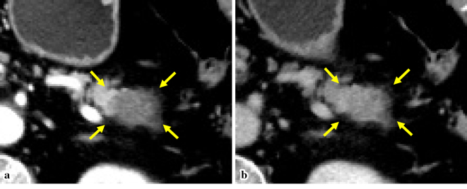
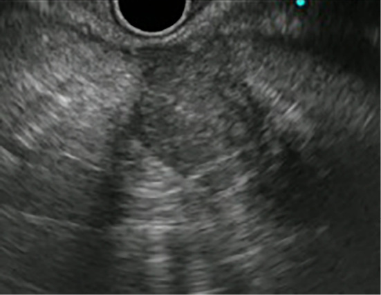
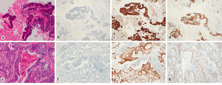
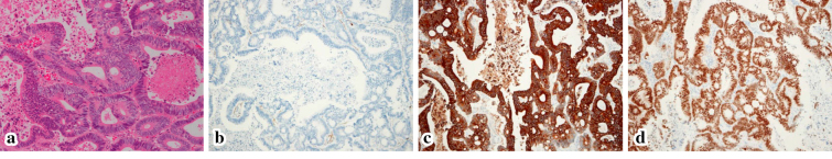
Pancreatic metastasis from colorectal cancer is rare, and there have been only a few reports of its preoperative diagnosis by endoscopic ultrasound-guided fine needle aspiration (EUS-FNA) with immunohistochemical staining. We herein describe the case of a 77-year-old woman in whom a solitary mass in the pancreatic tail was detected 11 years after rectal cancer resection. The patient also had a history of pulmonary tumor resection. We performed EUS-FNA and a histopathological examination showed adenocarcinoma with CD20+, CD7-, and CDX2+ (similar to her rectal cancer). EUS-FNA enabled a histopathological examination, including immunohistochemical staining, which helped to confirm the diagnosis of pancreatic and pulmonary metastasis from rectal cancer.
Figures
Similar articles
-
Utility of endoscopic ultrasound-guided fine-needle aspiration in the diagnosis and staging of colorectal carcinoma.Diagn Cytopathol. 2013 Dec;41(12):1031-7. doi: 10.1002/dc.21804. Epub 2011 Sep 19. Diagn Cytopathol. 2013. PMID: 21932358
-
Preoperative endoscopic ultrasound-guided fine needle aspiration does not impair survival of patients with resected pancreatic cancer.Gut. 2015 Jul;64(7):1105-10. doi: 10.1136/gutjnl-2014-307475. Epub 2015 Jan 9. Gut. 2015. PMID: 25575893
-
Needle tract seeding following endoscopic ultrasound-guided fine-needle aspiration for pancreatic cancer: a report of two cases.World J Surg Oncol. 2019 Aug 5;17(1):134. doi: 10.1186/s12957-019-1681-x. World J Surg Oncol. 2019. PMID: 31382964 Free PMC article.
-
Needle tract seeding recurrence of pancreatic cancer in the gastric wall with paragastric lymph node metastasis after endoscopic ultrasound-guided fine needle aspiration followed by pancreatectomy: a case report and literature review.BMC Gastroenterol. 2020 Jan 15;20(1):13. doi: 10.1186/s12876-020-1159-x. BMC Gastroenterol. 2020. PMID: 31941458 Free PMC article. Review.
-
Peritoneal dissemination of pancreatic cancer caused by endoscopic ultrasound-guided fine needle aspiration: A case report and literature review.World J Gastroenterol. 2021 Jan 21;27(3):294-304. doi: 10.3748/wjg.v27.i3.294. World J Gastroenterol. 2021. PMID: 33519143 Free PMC article. Review.
Cited by
-
A pancreatic metastasis from a colon carcinoma mimicking a primary tumor diagnosed by EUS-guided fine-needle biopsy.Endosc Ultrasound. 2023 Jan-Feb;12(1):162-163. doi: 10.4103/EUS-D-21-00186. Endosc Ultrasound. 2023. PMID: 35313422 Free PMC article. No abstract available.
-
Literature review of imaging, pathological diagnosis, and outcomes of metachronous lung and pancreatic metastasis of cecal cancer.World J Surg Oncol. 2022 Oct 17;20(1):341. doi: 10.1186/s12957-022-02797-7. World J Surg Oncol. 2022. PMID: 36253824 Free PMC article. Review.
-
Metachronous Pancreatic Metastasis from Rectal Cancer that Masqueraded as a Primary Pancreatic Cancer: A Rare and Difficult-to-Diagnose Metastatic Tumor in the Pancreas.Am J Case Rep. 2019 Nov 30;20:1781-1787. doi: 10.12659/AJCR.918669. Am J Case Rep. 2019. PMID: 31784503 Free PMC article.
References
-
- Z'graggen K, Fernández-del CC, Rattner DW, Sigala H, Warshaw AL. Metastases to the pancreas and their surgical extirpation. Arch Surg 133: 413-417, 1998. - PubMed
-
- Stankard CE, Karl RC. The treatment of isolated pancreatic metastases from renal cell carcinoma: a surgical review. Am J Gastroenterol 87: 1658-1660, 1992. - PubMed
-
- Inagaki H, Nakao A, Ando N, et al. . A case of solitary metastatic pancreatic cancer from rectal carcinoma: a case report. Hepatogastroenterology 45: 2413-2417, 1998. - PubMed
-
- Tanemura A, Mizuno S, Okura Y, et al. . Margin-negative limited resection of metastatic pancreatic tumors from rectal cancer preoperatively diagnosed by endoscopic ultrasound-guided fine-needle aspiration biopsies: report of two cases. Surg Today 44: 366-372, 2014. - PubMed
Publication types
MeSH terms
LinkOut - more resources
Full Text Sources
Other Literature Sources
Medical