Review
. 2017 Jun;16(6):400-423.
doi: 10.1038/nrd.2016.248. Epub 2017 Feb 3.

Targeting iron metabolism in drug discovery and delivery

Affiliations
Review

Targeting iron metabolism in drug discovery and delivery

Bart J Crielaard et al. Nat Rev Drug Discov. 2017 Jun.

Abstract

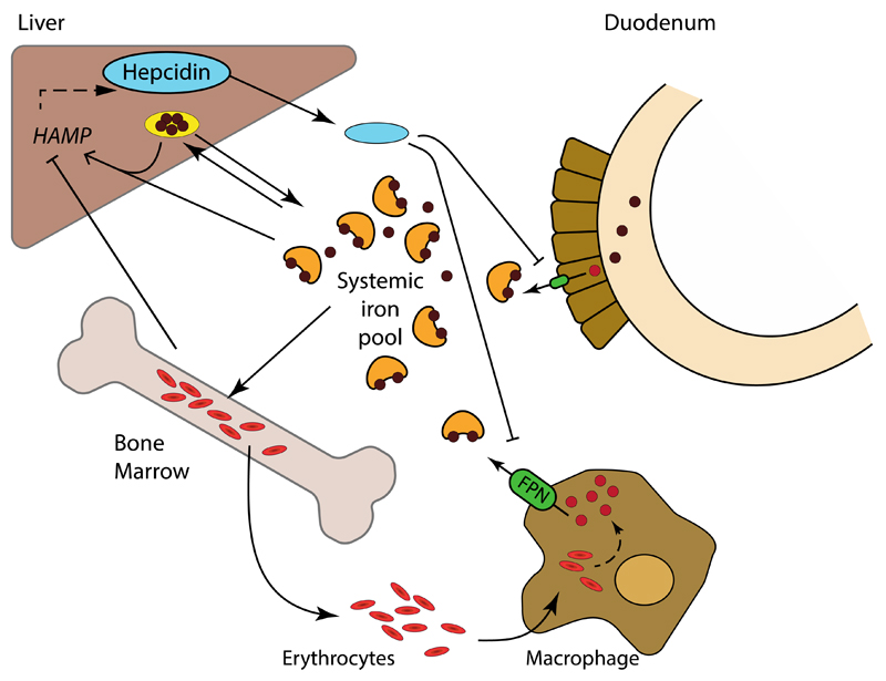
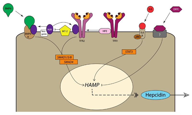
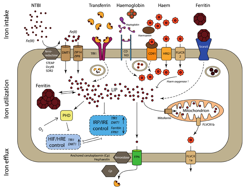
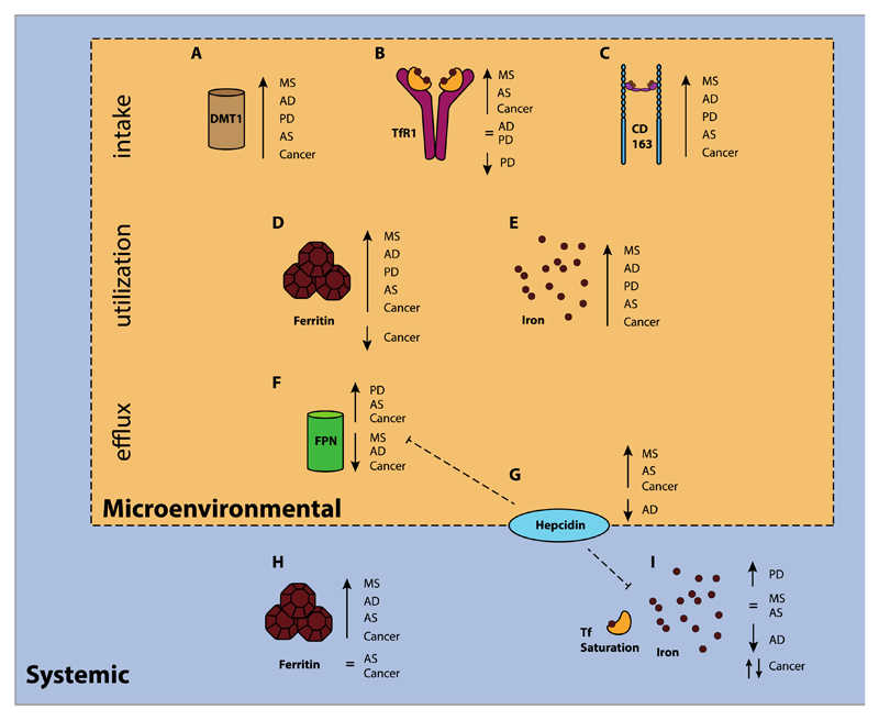
Iron fulfils a central role in many essential biochemical processes in human physiology; thus, proper processing of iron is crucial. Although iron metabolism is subject to relatively strict physiological control, numerous disorders, such as cancer and neurodegenerative diseases, have recently been linked to deregulated iron homeostasis. Consequently, iron metabolism constitutes a promising and largely unexploited therapeutic target for the development of new pharmacological treatments for these diseases. Several iron metabolism-targeted therapies are already under clinical evaluation for haematological disorders, and these and newly developed therapeutic agents are likely to have substantial benefit in the clinical management of iron metabolism-associated diseases, for which few efficacious treatments are currently available.


Conflict of interest statement

Competing financial interests

S.R. has restricted stocks in Merganser Biotech. S.R. is a consultant for Novartis Pharmaceuticals, Bayer Healthcare, Merganser Biotech and Keryx Pharmaceuticals. S.R. is a member of scientific advisory board of Merganser Biotech and Ionis Pharmaceuticals. T.L. is a member of scientific advisory board of Cristal Therapeutics B.V‥

Figures

Figure 1
Figure 1. Systemic iron metabolism.
Dietary iron is absorbed by duodenal enterocytes and added to the systemic iron pool upon export via ferroportin (FPN, green). The systemic iron pool, which under normal steady-state conditions is mainly bound to transferrin (orange), is primarily utilized for erythropoiesis. Excess systemic iron is stored as ferritin (yellow) in the liver, and to a lesser extent in other tissues. The iron utilized in senescent erythrocytes is recycled by macrophages and released, via ferroportin, back into the systemic iron pool. Ferroportin expression, and consequently systemic iron availability, is inhibited by hepcidin (blue), a peptide primarily produced by the liver. In turn, transcription of the gene encoding hepcidin, HAMP, is stimulated by the presence of iron and inhibited by erythropoietic demand.
Figure 2
Figure 2. Regulation of hepcidin expression in hepatocytes.
Bone morphogenetic protein 6 (BMP6) is produced by non-parenchymal cells in the liver in response to iron stores and flux. Binding of BMP6 to the BMP receptor complex on hepatocytes, in conjunction with hemojuvelin (HJV) results in downstream signalling via SMAD1/5/8 and SMAD4, which in turn stimulates HAMP transcription and expression of hepcidin. HJV is stabilized by neogenin, and cleaved by matriptase 2 (MT-2), allowing for fine-tuning of BMP6 signalling. SMAD signalling is additionally regulated by transferrin receptor 2 (TfR2), upon association with human hemochromatosis protein (HFE) and HJV. HFE also interacts with TfR1, where it competes with holotransferrin. Consequently, when iron supply is high, HFE is displaced from TfR1 allowing it to associate with TfR2, thereby increasing hepcidin expression via SMAD signalling. Under inflammatory conditions, interleukin 6 (IL-6) and related cytokines bind IL-6 receptor, resulting in JAK1/2-STAT3 activation and hepcidin expression to restrict iron availability. Finally, developing erythroid cells confer their iron requirements via secretion of erythroferrone (ERFE), which inhibits hepcidin expression, via a currently unknown receptor and signalling pathway, to stimulate iron uptake under circumstances of high erythropoietic demand.
Figure 3
Figure 3. Cellular iron trafficking pathways.
Iron intake. Non-transferrin bound iron (NTBI) can be transported directly (solid arrow) into the cell by DMT1, ZIP14 and ZIP8, after reduction of Fe(III) to Fe(II) by ferrireductases STEAP, DcytB or SDR2. Transferrin-bound iron is taken up via binding to TfR1 and endocytosis (dashed arrow) of the receptor, release and reduction of Fe(III) and transport into the cytoplasm via DMT1. Haemoglobin associates with haptoglobin to allows endocytosis by CD163, upon which haemoglobin is degraded, and haem transported into the cytosol. Systemic haem is scavenged by complexation with haemopexin and endocytosis via CD91. Haem can be directly transported, also from the endosome, into the cytosol by HRG1 and FLVCR2, where it is processed by haem oxygenase 1 and iron released as Fe(II). Scara5 internalizes ferritin that consists of light chains, after which iron is liberated and transported to the cytosol. Ferritin composed of heavy chains can be endocytosed by TfR1 (not depicted here). Iron utilization. The labile iron pool (LIP) comprises cytosolic Fe(II), which can be stored in cytoplasmic ferritin or utilized for biochemical processes. Most of these processes take place in the mitochondria, for which iron is supplied via mitoferrin, while FLVCR1b allows export of mitochondrially produced haem. The LIP controls the expression of TfR1, DMT1, ferritin and ferroportin via the posttranscriptional IRP/IRE regulatory system. Similarly, the transcriptional HIF/HRE regulatory system controls several intracellular processes, including the transcription of TfR1 and DMT1. HIF2α translation is inhibited by IRP1, while, conversely, IRP1 transcription is inhibited by HIF. HIF expression is regulated by PHD, which stimulates the degradation of HIFs under conditions of high oxygen. Iron egress. Fe(II) is exported by ferroportin, followed by oxidation to Fe(III) by the ferroxidases hephaestin or ceruloplasmin, and subsequent binding to transferrin. Intracellular haem can be directly exported via FLVCR1a. Not all pathways are present in all cells.
Figure 4
Figure 4. Up- and downregulation of several key players in non-haematological pathologies.
Schematic overview illustrating the microenvironmental and systemic changes in iron and iron metabolism-related proteins in selected pathologies in patients: multiple sclerosis (MS, sclerotic plaques), Alzheimer’s disease (AD, frontal cerebral cortex), Parkinson’s disease (PD, basal ganglia), atherosclerosis (AS, sclerotic plaques), and cancer (tumor tissue). A. Upregulation of DMT1 has been observed in MS (preclinical), AD (preclinical), PD, and cancer (colorectal). B. TfR1 upregulation has been found in MS, atherosclerosis, and cancer (colorectal, breast,, glioblastoma; normal TfR1 levels have been observed in AD,, while for PD normal levels, and low levels have been reported,. C. Increased levels of the macrophage-associated scavenger receptor CD163 have been found in affected tissues of patients with MS, AD, PD, AS, and cancer (breast, prostate, glioblastoma. D. Tissue iron stored as ferritin has found to be increased in MS, AD, PD, AS,, and for cancer both high levels (glioblastoma) and low levels have been reported (breast). E. Similarly, microenvironmental iron deposits have been observed in MS, AD,,, PD,,, AS, and cancer (colorectal). F. Upregulation of FPN has been demonstrated in tissues of patients with PD, AS, and cancer (colorectal, breast101), while low levels of FPN have been observed in MS (preclinical), AD and also in breast and prostate cancer. G. Hepcidin levels are not in all diseases consistent with (an inversed) FPN expression. High hepcidin concentrations have been found in MS (preclinical, tissue), AS (serum), breast cancer (tissue, and serum276) and prostate cancer (tissue), whereas a low level has been observed in the tissues of patients with AD. H. Increased systemic ferritin concentrations have found in the cerebrospinal fluid (CSF) of patients with MS, AD, and glioblastoma, as well as in the serum of patients with AS and breast cancer, although the levels of serum ferritin have also been found to be normal in patients with AS and non-skin cancers. I. Systemic iron levels measured by iron concentration and transferrin saturation have been quite inconsistent: higher than average levels have been found in a meta analysis in PD, normal levels have been found in MS and AS, and low levels have been observed in AD,. With regard to cancer, the association with systemic iron levels is quite unclear: women with high levels of serum iron were at higher risk for developing non-skin cancer, while men were at lower risk.
Figure 5
Figure 5. Proposed mechanisms for targeted drug delivery exploiting iron metabolism-associated cellular targets.
The natural ligand and the merits of its use as a delivery strategy are presented for each target. TfR1, CD163 and Scara5 allow endocytosis of drugs associated with their natural ligands. Subsequent lysosomal processing would then liberate the targeted drug, which then enters the cytosol by passive diffusion from the lysosome. This likely also occurs for haem-targeted constructs upon internalization by CD91, but possibly direct transport of the haem-linked drug into the cytosol by HRG1 and/or FLVCR2 could represent an alternative mechanism. Drug targeting to FPN, for example by using hepcidin, has not been explored until now, but forms an interesting approach for directing therapeutics to cell that overexpress FPN, such as some types of tumour cells. Tf, transferrin; Hb, haemoglobin; FPN, ferroportin

Similar articles

Cited by

References

    1. Rouault TA. Iron-sulfur proteins hiding in plain sight. Nat Chem Biol. 2015;11:442–445. - PubMed
    1. Hohenberger J, Ray K, Meyer K. The biology and chemistry of high-valent iron-oxo and iron-nitrido complexes. Nat Commun. 2012;3:720. - PubMed
    1. Ganz T. Systemic Iron Homeostasis. Physiol Rev. 2013;93:1721–1741. - PubMed
    1. Camaschella C. Iron-Deficiency Anemia. N Engl J Med. 2015;372:1832–1843. - PubMed
    1. Rivella S. β-thalassemias: paradigmatic diseases for scientific discoveries and development of innovative therapies. Haematologica. 2015;100:418–430. - PMC - PubMed