Review
. 2017 Jan 19:11:3.
doi: 10.3389/fnins.2017.00003. eCollection 2017.

Metal Dyshomeostasis and Their Pathological Role in Prion and Prion-Like Diseases: The Basis for a Nutritional Approach

Affiliations
Review

Metal Dyshomeostasis and Their Pathological Role in Prion and Prion-Like Diseases: The Basis for a Nutritional Approach

Mattia Toni et al. Front Neurosci. .

Abstract

Metal ions are key elements in organisms' life acting like cofactors of many enzymes but they can also be potentially dangerous for the cell participating in redox reactions that lead to the formation of reactive oxygen species (ROS). Any factor inducing or limiting a metal dyshomeostasis, ROS production and cell injury may contribute to the onset of neurodegenerative diseases or play a neuroprotective action. Transmissible spongiform encephalopathies (TSEs), also known as prion diseases, are a group of fatal neurodegenerative disorders affecting the central nervous system (CNS) of human and other mammalian species. The causative agent of TSEs is believed to be the scrapie prion protein PrPSc, the β sheet-rich pathogenic isoform produced by the conformational conversion of the α-helix-rich physiological isoform PrPC. The peculiarity of PrPSc is its ability to self-propagate in exponential fashion in cells and its tendency to precipitate in insoluble and protease-resistance amyloid aggregates leading to neuronal cell death. The expression "prion-like diseases" refers to a group of neurodegenerative diseases that share some neuropathological features with prion diseases such as the involvement of proteins (α-synuclein, amyloid β, and tau) able to precipitate producing amyloid deposits following conformational change. High social impact diseases such as Alzheimer's and Parkinson's belong to prion-like diseases. Accumulating evidence suggests that the exposure to environmental metals is a risk factor for the development of prion and prion-like diseases and that metal ions can directly bind to prion and prion-like proteins affecting the amount of amyloid aggregates. The diet, source of metal ions but also of natural antioxidant and chelating agents such as polyphenols, is an aspect to take into account in addressing the issue of neurodegeneration. Epidemiological data suggest that the Mediterranean diet, based on the abundant consumption of fresh vegetables and on low intake of meat, could play a preventive or delaying role in prion and prion-like neurodegenerative diseases. In this review, metal role in the onset of prion and prion-like diseases is dealt with from a nutritional, cellular, and molecular point of view.

Keywords: Alzheimer's disease; Mediterranean diet; Parkinson's disease; metal dyshomeostasis; prion; synuclein; synucleinopathies; transmissible spongiform encephalopathies.


Figures

Figure 1
Figure 1
Alignment of the α-syn amino acid sequences of representative species of teleost fish (Xiphophorus maculatus, accession number XP_005812724), amphibians (Xenopus laevis, NP_001080623), reptiles (Anolis carolinensis, XP_003221349), birds (Taeniopygia guttata, NP_001041718), and mammals (Homo sapiens, NP_001139526). Sequences were aligned with Clustal Omega (http://www.ebi.ac.uk/Tools/msa/clustalo/). Asterisks indicate identity of amino acids; double dots indicate amino acids with the same polarity or size; dots indicate semiconserved substitutions. Cu2+ binding sites are indicated by arrowheads, Cu+ binding regions are underlined, manganese, and iron binding sites are highlighted in gray and Ca2+ binding regions are double underlined. Negatively charged residues are indicated in bold characters. Circled P letter indicates residues whose phosphorylation increases the α-syn binding affinity for Cu2+, Pb2+, and Fe2+.
Figure 2
Figure 2
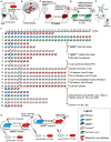
Schematic overview of the main evidence in support of the prion nature of human α-syn. (A) In vitro experiments, recombinant α-syn monomer can generate amylodogenic fibrils able to infect cultured cells. (B) In the idiopathic PD the formation of proteinaceous inclusion bodies begins in the dorsal motor nucleus of the vagus nerve and advances from there essentially upwards through susceptible regions of the medulla oblongata, pontine tegmentum, midbrain, and basal forebrain until it reaches the cerebral cortex (circled numbers refer to the synuclein progression in the CNS). (C) 12–22 years after transplantation into the striatum of individuals with PD, grafted nigral neurons developed α-syn positive LBs that stained positively also for ubiquitin providing evidence that the disease can propagate from host to graft cells. (D) Dopaminergic neurons extracted from the ventral mesencephalon of E12.5 C57BL/6 mice were grafted to the striatum of 6-week old transgenic mice overexpressing h α-syn. After 6 months, the grafted dopaminergic neurons showed putative intracellular h α-syn positive punctae demonstrating in vivo transfer of α-syn between host cells and grafted dopaminergic neurons. (E) Cell-produced α-syn is secreted via an exosomal calcium-dependent mechanism. The extracellular α-syn is taken up by cells through endocytosis and inside the cell interacts with intracellular α-syn forming dimers. (F) (1) Homozygous TgM83+/+ mice expressing A53T h α-syn in CNS neurons developed intracytoplasmic neuronal α-syn inclusions and a severe and complex motor impairment leading to paralysis and death starting from the 8th month; (2) hemizygous TgM83+/− mice developed the same symptoms between 22 and 28 months of age; (3) Tg mice that express wild type h α-syn developed no motor impairment and revealed normal neuropil staining pattern expected for the protein up to the age of 28 months; (4) TgM83+/+ mice inoculated with brain homogenates from sick 12 or 18 month-old TgM83+/+ mice showed the characteristic motor clinical signs of illness after 97 days post-inoculation (dpi); (5) bigenic Tg mice (M83+/−; Gfap-luc) inoculated with brain homogenates from spontaneously ill 10 month-old TgM83+/+ sample showed symptoms of synucleinopathies 160 dpi; (6) C57B1/6S Δα-syn mice (presenting a deletion of the α-syn locus) inoculated with brain homogenates from sick, 12 or 18 month-old TgM83+/+ mice, show no signs of disease and were still alive and healthy 14 months post-inoculation; (7) brain homogenate from healthy 2 month-old TgM83+/+ inoculated in TgM83+/+ mice did not induce an acceleration in the onset of α-syn pathology in TgM83+/+ mice; (8) Tg mice (M83+/−; Gfap-luc) inoculated with brain homogenate from two independent confirmed cases of MSA began to exhibit signs of neurologic illness, most commonly ataxia and circling behavior, at about 90 dpi; (9) WT C57BL6/C3H mice inoculated with synthetic pre-formed α-syn fibrils (PFF) obtained in vitro from recombinant mouse or h α-syn progressively developed α-syn cytoplasmatic accumulation at 30 dpi that evolved in dense perinuclear LB-like inclusions by 90 and 180 dpi; (10) α-syn−/− mice inoculated with PFF did not develop α-syn deposits; (11) no phosphorylated α-syn, ubiquitin or p62-positive pathology was observed in the brain of WT C57BL/6J mice inoculated with human or mouse α-syn monomer; (12) WT C57BL/6 mice inoculated with nigral LB-enriched fractions from post mortem PD brain resulted in progressive nigrostriatal neurodegeneration and diffusely α-syn accumulation within nigral neurons and anatomically interconnected regions by 4 up to 17 months post-inoculation; (13) WT C57BL/6 mice inoculated with nigral non-LB fractions from post mortem PD brain showed no nigrostriatal degeneration; (14) WT C57BL/6 mice inoculated with α-syn-immunodepleted nigral LB fractions from post mortem PD brain showed no phospho α-syn-positive cells and any evidence of pathology; (15) α-syn−/− C57Bl6Sv129 mice inoculated with nigral LB fractions from post mortem PD brain did not produce any α-syn pathology or evidence of nigrostriatal lesions; (16) WT C57BL/6J mice brain injected with DLB insoluble fraction from post mortem PD brain showed in the 50% of cases immunopositive structures for anti-phosphorylated α-syn at 15 months post injection (17–21) recombinant full length mouse fibrillar α-syn (mfib) or fibrillar human 21-140 α-syn (hfib) were injected in the hind limb muscle of 2 month-old TgM83+/+ (17), of TgM83+/− (18–19), of WT C3H/C57BL6 (20), or of α-syn−/− (21) mice. The injected mice died or had to be killed due to paralysis within 57–88 (TgM83+/+) or 121 (TgM83+/−) dpi. Mice also developed α-syn inclusion pathology that was nearly indistinguishable morphologically in anatomic distribution from that seen in aged (>8 month-old) untreated TgM83+/+ mice. Four of the seven mice that had sciatic nerve transection showed no motor deficits 200 dpi (19). On the contrary, WT and α-syn−/− mice developed no motor phenotype or α-syn pathology at 12 months post injection (20–21); (22) The injection of human Δ71–82 α-syn in TgM83+/+ mice resulted in delayed onset of disease (120 dpi) and incomplete penetrance of the pathology. (G) Inoculation with MSA brain homogenate in TgM83+/− mice caused CNS dysfunction and the accumulation of large aggregates of phosphorylated α-syn and widespread astrocytic gliosis with mean incubation periods of 106–143 dpi (primary transmission). The inoculations in TgM83+/− mice with brain homogenates from ill TgM83+/− mice previously inoculated with MSA showed a shorter incubation period (92–113) (secondary transmission). The brain homogenate from MSA patients and from both serially infected TgM83+/− mice were able to infect cultured cells. Figure drawings refers to the following references: (A; Desplats et al., ; Luk et al., ; Emmanouilidou et al., ; Nonaka et al., ; Volpicelli-Daley et al., ; Narkiewicz et al., 2014); (B; Braak et al., 2006); (C; Kordower et al., ; Li et al., , ; Kurowska et al., 2011); (D; Hansen et al., 2011); (E; Emmanouilidou et al., ; Hansen et al., 2011); [F: 1–3 (Giasson et al., 2002); 4, 6, 7 (Mougenot et al., 2012); 5, 8 (Watts et al., 2013); 9, 10 (Luk et al., 2012); 11, 16 (Masuda-Suzukake et al., 2013); 12–15 (Recasens and Dehay, 2014); 17–21 (Sacino et al., 2014)]; (G; Prusiner et al., 2015).

References

    1. Abeliovich A., Schmitz Y., Fariñas I., Choi-Lundberg D., Ho W. H., Castillo P. E., et al. . (2000). Mice lacking α-synuclein display functional deficits in the nigrostriatal dopamine system. Neuron 25, 239–252. 10.1016/S0896-6273(00)80886-7 - DOI - PubMed
    1. Acosta-Cabronero J., Betts M. J., Cardenas-Blanco A., Yang S., Nestor P. J. (2016). In vivo MRI mapping of brain iron deposition across the adult lifespan. J. Neurosci. 36, 364–374. 10.1523/JNEUROSCI.1907-15.2016 - DOI - PMC - PubMed
    1. Alcalay R. N., Gu Y., Mejia-Santana H., Cote L., Marder K. S., Scarmeas N. (2012). The association between Mediterranean diet adherence and Parkinson's disease. Mov. Disord. 27, 771–774. 10.1002/mds.24918 - DOI - PMC - PubMed
    1. Appel-Cresswell S., Vilarino-Guell C., Encarnacion M., Sherman H., Yu I., Shah B., et al. . (2013). Alpha-synuclein p.H50Q, a novel pathogenic mutation for Parkinson's disease. Mov. Disord. 28, 811–813. 10.1002/mds.25421 - DOI - PubMed
    1. Ayton S., Lei P. (2014). Nigral iron elevation is an invariable feature of Parkinson's disease and is a sufficient cause of neurodegeneration. Biomed Res. Int. 2014:581256. 10.1155/2014/581256 - DOI - PMC - PubMed