. 2017 Jan 19:11:7.
doi: 10.3389/fnins.2017.00007. eCollection 2017.

Effects of Systemic Metabolic Fuels on Glucose and Lactate Levels in the Brain Extracellular Compartment of the Mouse

Affiliations

Effects of Systemic Metabolic Fuels on Glucose and Lactate Levels in the Brain Extracellular Compartment of the Mouse

Alexandria Béland-Millar et al. Front Neurosci. .

Abstract

Classic neuroenergetic research has emphasized the role of glucose, its transport and its metabolism in sustaining normal neural function leading to the textbook statement that it is the necessary and sole metabolic fuel of the mammalian brain. New evidence, including the Astrocyte-to-Neuron Lactate Shuttle hypothesis, suggests that the brain can use other metabolic substrates. To further study that possibility, we examined the effect of intraperitoneally administered metabolic fuels (glucose, fructose, lactate, pyruvate, ß-hydroxybutyrate, and galactose), and insulin, on blood, and extracellular brain levels of glucose and lactate in the adult male CD1 mouse. Primary motor cortex extracellular levels of glucose and lactate were monitored in freely moving mice with the use of electrochemical electrodes. Blood concentration of these same metabolites were obtained by tail vein sampling and measured with glucose and lactate meters. Blood and extracellular fluctuations of glucose and lactate were monitored for a 2-h period. We found that the systemic injections of glucose, fructose, lactate, pyruvate, and ß-hydroxybutyrate increased blood lactate levels. Apart for a small transitory rise in brain extracellular lactate levels, the main effect of the systemic injection of glucose, fructose, lactate, pyruvate, and ß-hydroxybutyrate was an increase in brain extracellular glucose levels. Systemic galactose injections produced a small rise in blood glucose and lactate but almost no change in brain extracellular lactate and glucose. Systemic insulin injections led to a decrease in blood glucose and a small rise in blood lactate; however brain extracellular glucose and lactate monotonically decreased at the same rate. Our results support the concept that the brain is able to use alternative fuels and the current experiments suggest some of the mechanisms involved.

Keywords: astrocytes; beta-hydroxybutyrate; blood-brain barrier; brain metabolism; fructose; galactose; insulin; pyruvate.


Figures

Figure 1
Figure 1
Histology and Stereotaxic Location of Electrodes. On the right is a picture of a 14 μm mouse brain slice taken following the implantation of an electrode in the primary motor cortex (M1). For comparison, on the left is a picture from the Franklin and Paxinos (2008) Mouse Brain Atlas used to determine stereotaxic coordinates and confirm electrode placement in the primary motor cortex.
Figure 2
Figure 2
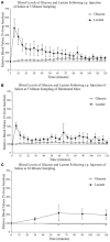
Relative blood concentration of glucose and lactate following a 0.9% i.p. injection of saline (A) at 5 min sampling interval in naïve mice (n = 4); (B) at 5 min sampling interval in habituated mice (n = 4); (C) and at 30 min sampling interval in naïve mice (n = 4). Values are expressed as a percent of their baseline value (taken prior to injection) and error bars represent the standard error of the mean.
Figure 3
Figure 3
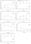
Relative blood concentration of glucose and lactate following a 2 g/kg i.p. injection of (A) glucose (n = 4); (B) fructose (n = 4); (C) lactate (n = 4); (D) pyruvate (n = 4); (E) β–hydroxybutyrate (n = 4); (F) galactose (n = 4). (G) Relative blood concentration of glucose and lactate following a 1.2 I.U. i.p. injection of insulin (n = 4). Values are expressed as a percent of their baseline value (taken prior to injection) and error bars represent the standard error of the mean. * = significant difference when compared to control (saline) values (p < 0.05) & = significant difference when compared to baseline (p < 0.05).
Figure 4
Figure 4
Relative extracellular concentration of glucose and lactate following a (A) 0.9% i.p. injection of saline (n = 4), a 2 g/kg i.p. injection of (B) glucose (n = 4); (C) fructose (n = 6); (D) lactate (n = 10); (E) pyruvate (n = 6); (F) β–hydroxybutyrate (n = 5); (G) galactose (n = 6), as well as (H) a 1.2 I.U. i.p. injection of insulin (n = 7). Values are expressed as a percent of their baseline value (taken prior to injection) and error bars represent the standard error of the mean. The thin horizontal lines below each graph represent significant tests for extracellular glucose values while the thicker horizontal line indicates significant tests for extracellular lactate values (p ≤ 0.05). Significant tests for comparisons between saline values are indicated separately from the comparison with baseline values.

References

    1. Abel E. L. (1994). A further analysis of physiological changes in rats in the forced swim test. Physiol. Behav. 56, 795–800. 10.1016/0031-9384(94)90245-3 - DOI - PubMed
    1. Abi-Saab W. M., Maggs D. G., Jones T., Jacob R., Srihari V., Thompson J., et al. . (2002). Striking differences in glucose and lactate levels between brain extracellular fluid and plasma in conscious human subjects: effects of hyperglycemia and hypoglycemia. J. Cereb. Blood Flow Metab. 22, 271–279. 10.1097/00004647-200203000-00004 - DOI - PubMed
    1. Adelman R. C., Spolter P. D., Weinhouse S. (1966). Dietary and hormonal regulation of enzymes of fructose metabolism in rat liver. J. Biol. Chem. 241, 5467–5472. - PubMed
    1. Adolphs R., Tranel D., Hamann S., Young A. W., Calder A. J., Phelps E. A., et al. . (1999). Recognition of facial emotion in nine individuals with bilateral amygdala damage. Neuropsychologia 37, 1111–1117. 10.1016/S0028-3932(99)00039-1 - DOI - PubMed
    1. Akiyoshi H., Iwamoto M., Nakaya Y. (1999). Lactate stimulates insulin secretion without blocking the K+ channels in HIT-T15 insulinoma cells. Horm. Metab. Res. 31, 257–261. 10.1055/s-2007-978728 - DOI - PubMed

LinkOut - more resources