Review
. 2017:2017:2752876.
doi: 10.1155/2017/2752876. Epub 2017 Jan 3.

Ultrasound-Guided Quadratus Lumborum Block: An Updated Review of Anatomy and Techniques

Affiliations
Review

Ultrasound-Guided Quadratus Lumborum Block: An Updated Review of Anatomy and Techniques

Hironobu Ueshima et al. Biomed Res Int. 2017.

Abstract

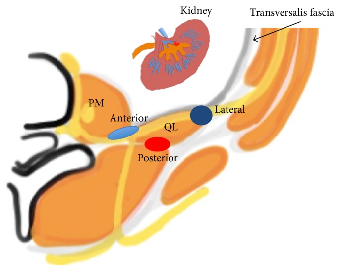
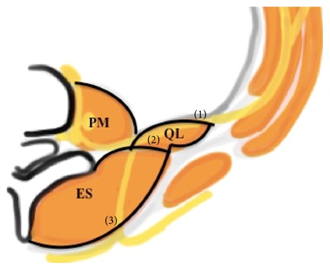
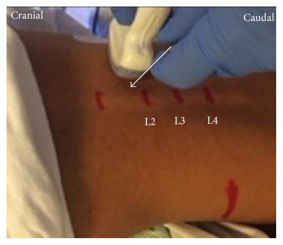
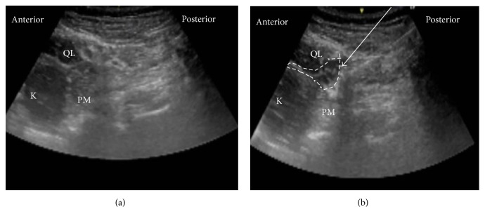
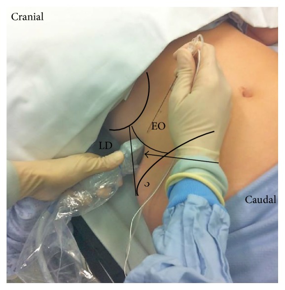
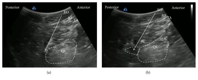
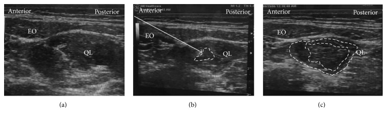
Purpose of Review. Since the original publication on the quadratus lumborum (QL) block, the technique has evolved significantly during the last decade. This review highlights recent advances in various approaches for administering the QL block and proposes directions for future research. Recent Findings. The QL block findings continue to become clearer. We now understand that the QL block has several approach methods (anterior, lateral, posterior, and intramuscular) and the spread of local anesthetic varies with each approach. In particular, dye injected using the anterior QL block approach spread to the L1, L2, and L3 nerve roots and within psoas major and QL muscles. Summary. The QL block is an effective analgesic tool for abdominal surgery. However, the best approach is yet to be determined. Therefore, the anesthetic spread of the several QL blocks must be made clear.


Conflict of interest statement

The authors declare that there is no conflict of interests.

Figures

Figure 1
Figure 1
Anatomic view of quadratus lumborum (QL) block (anterior, lateral, and posterior). The lateral QL block injects the local anesthetic at the lateral to the QL muscle. The posterior QL block injects the local anesthetic at the posterior to the QL muscle. The anterior QL block injected the local anesthetic between the PM muscle and the QL muscle. QL: quadratus lumborum muscle, PM: psoas major muscle, and gray line: transversalis fascia.
Figure 2
Figure 2
Anatomic view of the thoracolumbar fascia (TLF). The TLF is divided into 3 layers (anterior (1), middle (2), and posterior (3)). QL: quadratus lumborum, ES: erector spinae, LD: latissimus dorsi, and PM: psoas major.
Figure 3
Figure 3
Probe position for anterior QLB. The convex probe was vertically attached above the iliac crest.
Figure 4
Figure 4
Ultrasound images of anterior QLB. (a) Preinjection and (b) postinjection. QL: quadratus lumborum, PM: psoas muscle, white arrow: needle trajectory, and white dotted line: spread of local anesthetic.
Figure 5
Figure 5
Probe position for subcostal QL block. A low-frequency convex probe is placed with a transverse, oblique, and paramedian orientation approximately 3 cm lateral to the L2 spinous process.
Figure 6
Figure 6
Ultrasound images of subcostal QL block. (a) Preinjection and (b) postinjection. QL: quadratus lumborum, PM: psoas muscle, white arrow: needle trajectory, and white dotted line: spread of local anesthetic.
Figure 7
Figure 7
Lateral QL block. A high-frequency linear probe was attached in the area of the triangle of Petit. EO: external abdominal oblique; LD: latissimus dorsi; black arrow: the triangle of Petit.
Figure 8
Figure 8
Ultrasound images of lateral QLB. (a) Preinjection and (b) postinjection. EO: external oblique muscle, IO: internal oblique muscle, TA: transversus abdominis, QL: quadratus lumborum, white arrow: needle trajectory, and white dotted line: spread of local anesthetic.
Figure 9
Figure 9
Ultrasound images of posterior QLB. (a) Preinjection and (b) postinjection. EO: external oblique muscle, IO: internal oblique muscle, TA: transversus abdominis, QL: quadratus lumborum, white arrow: needle trajectory, and white dotted line: spread of local anesthetic.
Figure 10
Figure 10
Ultrasound images of intramuscular QLB. (a) Preinjection, (b) test injection, and (c) postinjection. EO: external oblique muscle, QL: quadratus lumborum, white arrow: needle trajectory, and white dotted line: spread of local anesthetic within (b) or in between (c).

References

    1. Blanco R. TAP block under ultrasound guidance: the description of a ‘non pops technique’. Regional Anesthesia and Pain Medicine. 2007;32(supplement 1):p. 130. - PubMed
    1. Kadam V. R. Ultrasound-guided quadratus lumborum block as a postoperative analgesic technique for laparotomy. Journal of Anaesthesiology Clinical Pharmacology. 2013;29(4):550–552. doi: 10.4103/0970-9185.119148. - DOI - PMC - PubMed
    1. Blanco R., Ansari T., Girgis E. Quadratus lumborum block for postoperative pain after caesarean section: a randomised controlled trial. European Journal of Anaesthesiology. 2015;32(11):812–818. doi: 10.1097/eja.0000000000000299. - DOI - PubMed
    1. Chakraborty A., Goswami J., Patro V. Ultrasound-guided continuous quadratus lumborum block for postoperative analgesia in a pediatric patient. A & A Case Reports. 2015;4(3):34–36. doi: 10.1213/xaa.0000000000000090. - DOI - PubMed
    1. Hebbard P. D. Transversalis fascia plane block, a novel ultrasound-guided abdominal wall nerve block. Canadian Journal of Anesthesia. 2009;56(8):618–620. doi: 10.1007/s12630-009-9110-1. - DOI - PubMed

MeSH terms

Substances

LinkOut - more resources