

Paraquat intoxication and associated pathological findings in three dogs in South Africa
- PMID: 28155296
- PMCID: PMC6138077
- DOI: 10.4102/jsava.v87i1.1352
Paraquat intoxication and associated pathological findings in three dogs in South Africa
Abstract
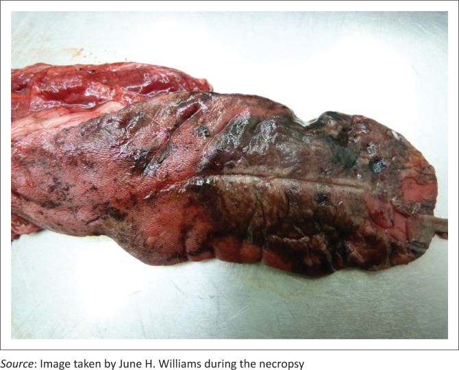
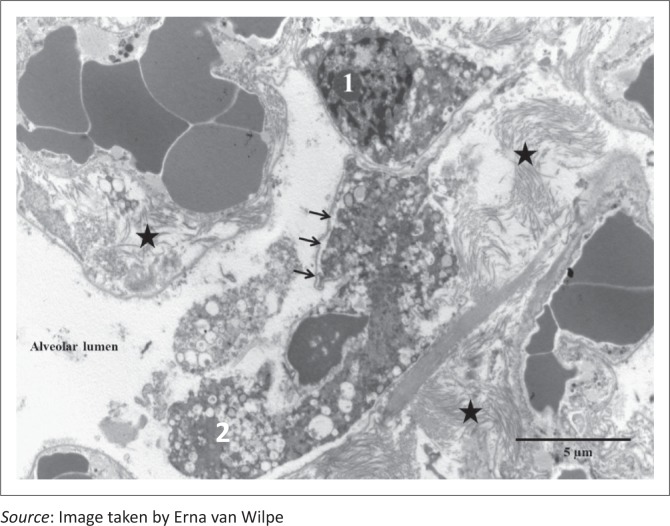
Paraquat is a bipyridylium non-selective contact herbicide commonly used worldwide. When ingestion occurs by humans and animals either accidentally, intentionally or maliciously, paraquat selectively accumulates in the lungs resulting in the production of oxygen-free radicals, causing membrane damage and cell death. Intoxicated subjects typically show progressive and fatal pulmonary haemorrhage, collapse and oedema. In individuals surviving the acute phase, pulmonary fibrosis develops. Gastrointestinal-, renal- and central nervous system clinical signs may also occur. Owing to the lack of effective treatment and absence of an antidote, the prognosis is poor. The clinical presentation, clinicopathological findings and treatment are briefly described of three dogs from one South African household, intoxicated with paraquat. Macroscopic and microscopic lesions in one dog that was necropsied, as well as pulmonary ultrastructure are detailed and illustrated for academic reference. All dogs presented with tachypnoea and dyspnoea 2-3 days after accidental paraquat ingestion. Treatment was aimed at reducing gastrointestinal absorption, enhancing elimination by diuresis and avoiding further oxidative damage by administration of antioxidants. All dogs, however, became progressively hypoxic despite treatment and were euthanised. Paraquat toxicity should be a differential diagnosis in dogs with unexplained progressive respiratory and gastrointestinal signs and renal failure. The local veterinary profession should be aware of accidental or intentional paraquat toxicity of animals. Existing literature, variations possible in canine clinical signs, measured parameters, lesions, as well as possible treatments, promising experimental antidotes and management options are discussed.
Conflict of interest statement
The authors declare that they have no financial or personal relationship(s) that may have inappropriately influenced them in writing this article.
Figures


References
-
- Bischoff K., Brizzee-Buxton B., Gatto N., Edwards W.C., Stair E.L. & Logan C, 1998, ‘Malicious paraquat poisoning in Oklahoma dogs’, Veterinary and Human Toxicology 40(3), 151–153. - PubMed
-
- Caswell J.L. & Williams K.J, 2007, ‘Respiratory system’, in Maxie M.G. (ed.), Jubb, Kennedy and Palmer’s pathology of domestic animals, 5th edn, pp. 574–575, Saunders Elsevier, London.
Publication types
MeSH terms
Substances
LinkOut - more resources
Full Text Sources
Other Literature Sources