. 2017 Feb 3:7:41807.
doi: 10.1038/srep41807.

New perspectives on corpora amylacea in the human brain

Affiliations

New perspectives on corpora amylacea in the human brain

Elisabet Augé et al. Sci Rep. .

Abstract

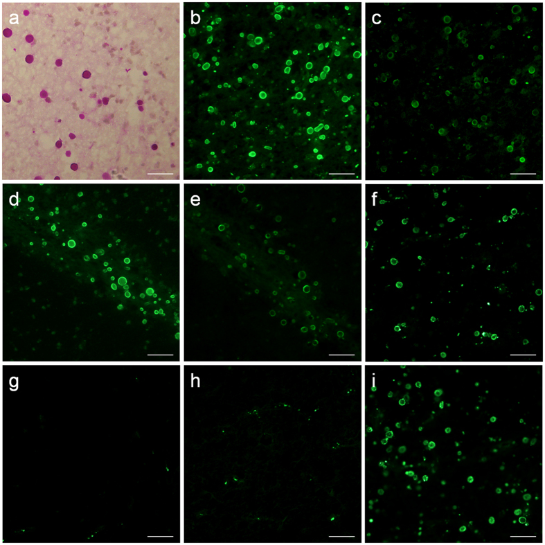
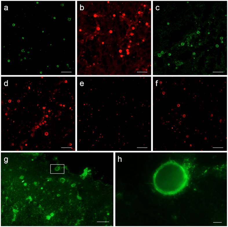
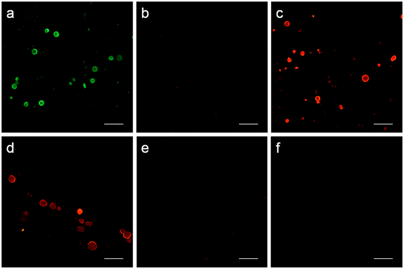
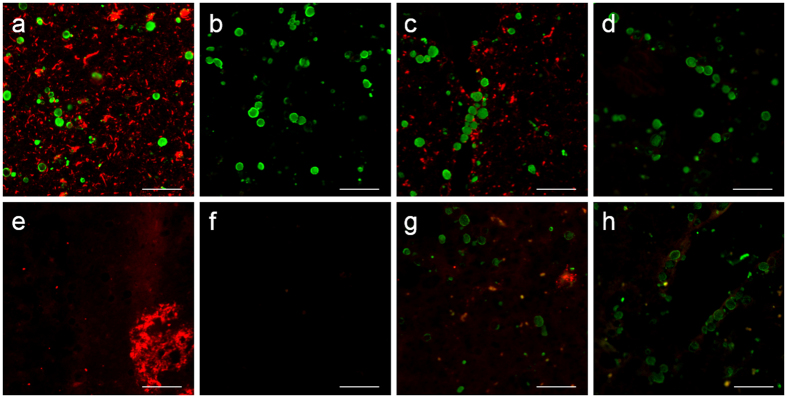
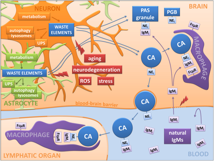
Corpora amylacea are structures of unknown origin and function that appear with age in human brains and are profuse in selected brain areas in several neurodegenerative conditions. They are constituted of glucose polymers and may contain waste elements derived from different cell types. As we previously found on particular polyglucosan bodies in mouse brain, we report here that corpora amylacea present some neo-epitopes that can be recognized by natural antibodies, a certain kind of antibodies that are involved in tissue homeostasis. We hypothesize that corpora amylacea, and probably some other polyglucosan bodies, are waste containers in which deleterious or residual products are isolated to be later eliminated through the action of the innate immune system. In any case, the presence of neo-epitopes on these structures and the existence of natural antibodies directed against them could become a new focal point for the study of both age-related and degenerative brain processes.


Conflict of interest statement

The authors declare no competing financial interests.

Figures

Figure 1
Figure 1. CA contain neo-epitopes that can be recognized by plasma IgMs.
A representative image of PAS staining illustrating the presence of CA in a hippocampal brain section from an AD donor is shown in (a). Representative images from brain sections from an AD donor (b and c) and a control donor (d and e) immunostained with two different human blood plasma in the first incubation and anti-human IgM (μ chain specific) conjugated to a fluorochrome in the second incubation (b and d correspond to one plasma donor, and c and e to the other donor). As can be observed, CA were positively stained in all cases. If commercial purified human IgM antibodies were used in the first incubation, CA were also stained (f). Negative staining was observed when BB (g) and the Fc fragment of human IgM (h) were used in the first incubation, instead of human IgMs. Previous incubation with an Fc receptor blocker did not block the positive staining obtained when commercial purified human IgM was used in the first incubation (i). The last four representative images (f–i) were obtained from the same AD donor. Scale bar: 50 μm.
Figure 2
Figure 2. The IgMs that recognize CA neo-epitopes are natural antibodies.
Representative images from an AD donor showing the positive staining of CA when using goat (a), rat (b), rabbit (c) and mouse (d) sera in the first incubation and an appropriate fluorochrome-labeled anti-IgM antibody in the second. Negative staining was obtained when BB was used in the first incubation instead of the different mammal sera and a representative image obtained when an anti-mouse IgM was used in the second incubation is shown (e). A representative image from the same AD donor showing the positive staining of CA with sera from SOPF mice is shown (f). Purified IgMs from umbilical cord sera positively stained the CA of an AD patient and a representative image is shown in (g). (h) Inset in (g). Scale bar (a–g) 50 μm; scale bar (h) 5 μm.
Figure 3
Figure 3. IgMs can produce false positive staining of CA.
Representative images of brain sections from an AD patient stained with JJ319 mouse IgG1 primary antibody and: a secondary fluorochrome-labeled antibody against heavy and light chains of mouse IgG (a) a secondary fluorochrome-labeled antibody against the γ1 chain of mouse IgG (b) and a secondary fluorochrome-labeled antibody against the μ chain of mouse IgM (c). As can be observed, CA are not stained when the secondary antibody against the γ1 chain of mouse IgG is used. When purified IgMs or purified IgGs from JJ319 antibody were used in the first incubation (d and e respectively) and a fluorochrome-labeled antibody against the μ chain was used in both cases in the second incubation, positive staining of CA was obtained when using purified IgMs while it was negative when using purified IgGs. Negative staining of CA was also obtained when purified IgGs were incubated followed by a secondary antibody against heavy and light chains of mouse IgG (f). It can therefore be concluded that only IgM bind to the CA and that the staining when using a secondary antibody against heavy and light chains of mouse IgG is just a cross-reaction with IgMs. Scale bar: 50 μm.
Figure 4
Figure 4. CA do not contain tau protein or amyloid–β peptides.
Representative images obtained from brain samples from an AD donor stained with: anti-tau clone tau 5 (a) pre-adsorbed anti-tau clone tau 5 (b) anti-tau clone tau 5E2 (c) and pre-adsorbed anti-tau clone tau 5E2 (d), followed by both fluorochrome-labeled secondary antibodies directed against the μ chain of mouse IgM (green) and against the γ1 chain of mouse IgG (red). When using non-pre-adsorbed antibodies, CA were positively stained with the contaminant IgMs contained in the anti-tau antibodies (green) and neurofibrillary tangles and neurons were stained with the anti-tau IgG1 (red) (a and c). When the antibodies were pre-adsorbed with tau protein, the staining of the neuro fibrillary tangles and neurons disappeared while that of CA by contaminant IgMs remained (b and d). Representative images obtained from brain samples from an AD donor stained with: the 6E10 antibody (e) pre-adsorbed 6E10 (f) the 12F4 antibody (g) and pre-adsorbed 12F4 (h), followed by both fluorochrome-labeled secondary antibodies directed against the μ chain of mouse IgM (green) and against the γ1 chain of mouse IgG (red). When using non-pre-adsorbed anti-Aβ IgG1 antibodies, amyloid plaques appeared positively stained with IgGs (red) (e and g). CA did not stain after 6E10 staining because this antibody did not contain contaminant IgM (e) while they appeared stained with 12F4 (green), denoting contamination of IgMs in this antibody (g). When the antibodies were pre-adsorbed with Aβ fragments, the staining of the amyloid plaques disappeared while that of CA by contaminant IgMs remained (h). Scale bar: 50 μm.
Figure 5
Figure 5. Schematic integrative proposal.
Waste elements generated from neurons, astrocytes, oligodendrocytes and other brain cells are removed intracellularly via the ubiquitin proteasome system (UPS) or via autophagic processes linked to lysosomal digestion. However, some of these waste elements can be found in polyglucosan bodies (PGBs) like CA in human brains or PAS granules in mouse brains, mainly when the production of waste elements is strengthened by factors such as aging, neurodegenerative disorders, reactive oxygen species or cellular stress. These PGBs contain some neo-epitopes (NEi) that are targeted by natural antibodies of the IgM type (IgMi). Since in physiological conditions the blood –brain barrier prevents access of IgMs to the brain parenchyma, the linkage between IgMs and CA is produced outside the brain after CA extrusion or inside the brain if the barrier is disrupted, thus allowing the passage of the IgMs. Macrophages, which contain specific receptors for the activated IgMs (FcμR), can subsequently phagocytize them. The new relationship shown here between PGBs and the natural immune system suggests that these PGBs could be waste containers ready to be eliminated by the action of this predetermined immune system.

References

    1. Cavanagh J. B. Corpora-amylacea and the family of polyglucosan diseases. Brain Res. Rev. 29, 265–295 (1999). - PubMed
    1. Rohn T. T. Corpora amylacea in neurodegenerative diseases: cause or effect? Int. J. Neurol. Neurother. 2, 31 (2015). - PMC - PubMed
    1. Pirici D. & Margaritescu C. Corpora amylacea in aging brain and age-related brain disorders. J. Aging Gerontol. 2, 33–57 (2014).
    1. Ferraro A. & Damon L. A. The histogenesis of amyloid bodies in the central nervous system. Arch. Pathol. 12, 229–244 (1931).
    1. Sakai M., Austin J., Witmer F. & Trueb L. Studies of corpora amylacea: Isolation and preliminary characterization by chemical and histochemical techniques. Arch. Neurol. 21, 526–544 (1969). - PubMed

Publication types

MeSH terms