. 2017 Feb 2;9(2):27.
doi: 10.3390/v9020027.

Myxoma Virus dsRNA Binding Protein M029 Inhibits the Type I IFN-Induced Antiviral State in a Highly Species-Specific Fashion

Affiliations

Myxoma Virus dsRNA Binding Protein M029 Inhibits the Type I IFN-Induced Antiviral State in a Highly Species-Specific Fashion

Masmudur M Rahman et al. Viruses. .

Abstract

Myxoma virus (MYXV) is Leporipoxvirus that possesses a specific rabbit-restricted host tropism but exhibits a much broader cellular host range in cultured cells. MYXV is able to efficiently block all aspects of the type I interferon (IFN)-induced antiviral state in rabbit cells, partially in human cells and very poorly in mouse cells. The mechanism(s) of this species-specific inhibition of type I IFN-induced antiviral state is not well understood. Here we demonstrate that MYXV encoded protein M029, a truncated relative of the vaccinia virus (VACV) E3 double-stranded RNA (dsRNA) binding protein that inhibits protein kinase R (PKR), can also antagonize the type I IFN-induced antiviral state in a highly species-specific manner. In cells pre-treated with type I IFN prior to infection, MYXV exploits M029 to overcome the induced antiviral state completely in rabbit cells, partially in human cells, but not at all in mouse cells. However, in cells pre-infected with MYXV, IFN-induced signaling is fully inhibited even in the absence of M029 in cells from all three species, suggesting that other MYXV protein(s) apart from M029 block IFN signaling in a speciesindependent manner. We also show that the antiviral state induced in rabbit, human or mouse cells by type I IFN can inhibit M029-knockout MYXV even when PKR is genetically knocked-out, suggesting that M029 targets other host proteins for this antiviral state inhibition. Thus, the MYXV dsRNA binding protein M029 not only antagonizes PKR from multiple species but also blocks the type I IFN antiviral state independently of PKR in a highly species-specific fashion.

Keywords: Myxoma virus; M029; Poxvirus; dsRNA binding protein; type I IFNs, antiviral state.


Conflict of interest statement

The authors declare no conflict of interest.

Figures
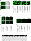
Figure 1
Figure 1
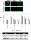
Myxoma virus (MYXV) is able to inhibit the type I interferon (IFN)-induced antiviral state completely in rabbit cells, partially in human cells and poorly in mouse cells. RK13 cells were individually treated with uIFNα, hIFNβ or rIFN containing media for 18 h and (A) infected with vesicular stomatitis virus (VSV)-GFP at a multiplicity of infection (MOI) of 1.0, fluorescence images were taken 48 h post-infection (hpi) and a representative image is shown; scale bars (100 µm); (B) infected with the wild-type (WT)-MYXV at an MOI of 0.01 and fluorescence images were taken 48 hpi; (C) infected with the wild-type (WT)-MYXV at an MOI of 5.0 and then cells were collected at 1 h (adsorption), 24 h and 48 hpi for virus titration; (D) Human GM02504 primary fibroblasts or HeLa cells were treated with uIFNα or hIFNβ containing media for 18 h and infected with the WT-MYXV at an MOI of 0.01 and fluorescence images were taken 48 h after infection; (E) GM02504 cells were mock or treated with uIFNα or hIFNβ containing media for 18 h and infected with the WT-MYXV at an MOI of 5.0 and then cells were collected at 1 h (adsorption), 24 h and 48 h after infection for virus titration; (F) Mouse embryo fibroblast (MEF) cells were treated with uIFNα or mIFNβ containing media for 18 h and infected with the WT-MYXV at an MOI of 1.0 and fluorescence images were taken 48 h after infection; (G) MEF cells were mock or treated with uIFNα or mIFNβ containing media for 18 h and infected with the WT-MYXV at an MOI of 5.0 and then cells were collected at 1 h (adsorption), 24 h and 48 hpi for virus titration. The virus titers were determined in triplicate following serial dilution onto RK13 cells. Statistics are relative to untreated virus titer after 24 h or 48 h infection. ** p < 0.01 *** p < 0.001; (H) Table showing log10 changes in virus titer after different type I IFN treatment compare to the untreated samples at 24 h and 48 h in RK13, GM02504 and MEFs.
Figure 2
Figure 2
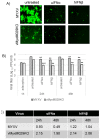
M029 is required for the complete inhibition of the type I IFN-induced antiviral state in rabbit RK13 cells. (A) RK13 cells were treated with uIFNα, hIFNβ or rIFN containing media for 18 h and infected with the vMyxM029KO virus at an MOI of 0.1 and fluorescence images were taken 48 hpi; (B) RK13 cells were mock or treated with uIFNα, hIFNβ or rIFN containing media for 18 h and infected with the vMyxM029KO virus at an MOI of 5.0 and then cells were collected at 1 h (adsorption), 24 h and 48 hpi for virus titration. The virus titers were determined in triplicate following serial dilution onto permissive RK13-E3 cells. Statistics are relative to untreated virus titer after 24 h or 48 hpi. ** p < 0.01 *** p < 0.001; (C) Table showing log10 changes in virus titer after different type I IFN treatment compare to the untreated samples at 24 h and 48 h in RK13.
Figure 3
Figure 3
Loss of protein kinase R (PKR) can rescue M029-minus MYXV replication in mouse cells but not overcome sensitivity to the mouse type I IFN-induced antiviral state. (A) MEF PKR-/- cells were treated with uIFNα, or mIFNβ containing media for 18 h and infected with the WT-MYXV or vMyxM029KO virus at an MOI of 0.1 and fluorescence images were taken 48 hpi; (B) MEF PKR-/- cells were mock or treated with uIFNα or mIFNβ containing media for 18 h and infected with the WT-MYXV or vMyxM029KO viruses at an MOI of 5.0 and then cells were collected at 1 h (adsorption), 24 h and 48 hpi for virus titration. The virus titers were determined in triplicate following serial dilution onto permissive RK13-E3 cells. Statistics are relative to virus titer after 24 h or 48 h infection. ** p < 0.01 *** p < 0.001; (C) Table showing log10 changes in virus titer after different type I IFN treatment compare to the untreated samples at 24 h and 48 h in MEF PKR-/- cells.
Figure 4
Figure 4
CRISPR-mediated PKR knockout in HeLa cells can rescue vMyxM029KO virus replication. (A) Generation of PKR knockout cells by CRISPR/CAS9 technology. HeLa cells were transfected with the CRISPR/Cas9 plasmids as described in Materials and Methods. Cells from different colonies (represented as KO) expanded, treated with hIFNβ for 18 h, lysed, and total proteins were resolved by SDS-PAGE and analyzed by Western blotting to detect endogenous PKR, IFN-stimulated gene 15 (ISG15) and actin as a loading control. Colony 2 (KO:2) was selected for further experiments and labeled as HeLa PKR-/- cells; (B) vMyxM029KO virus is able to replicate in the HeLa PKR-/- colonies. HeLa ctrl or HeLa PKR-/- colonies were infected with vMyxM029KO virus and fluorescence images were taken 48 hpi; (C) Single step growth curves of MYXV infection in HeLa ctrl and HeLa PKR-/- cells. The indicated cells were infected with WT-MYXV or vMyxM029KO at an MOI of 5, and then cells were collected at 1, 6, 18, 30 and 48 hpi. The virus titers were determined in triplicate following serial dilution onto RK13-E3 cells. Data are representative of three independent experiments.
Figure 5
Figure 5
M029 is required for partial inhibition of the type I IFN-induced antiviral state in human cells; (A) HeLa PKR-/- cells were treated with uIFNα or hIFNβ containing media for 18 h and infected with the WT-MYXV or vMyxM029KO viruses at an MOI of 0.01 and fluorescence images were taken 48 hpi; (B) HeLa PKR-/- cells were mock or treated with uIFNα, hIFNβ containing media for 18 h and infected with the WT-MYXV or vMyxM029KO viruses at an MOI of 5.0 and then cells were collected at 1 h (adsorption), 24 h and 48 hpi for virus titration. The virus titers were determined in triplicate following serial dilution onto permissive RK13-E3 cells. Statistics are relative to untreated virus titer after 24 h or 48 h infection. ** p < 0.01 *** p < 0.001; (C) Table showing log10 changes in virus titer after different type I IFN treatment compare to the untreated samples at 24 h and 48 h in HeLa PKR-/- cells.
Figure 6
Figure 6
Knockdown of PKR cannot rescue M029-minus MYXV sensitivity to the type I IFN-induced antiviral state in rabbit RK13 cells. RK13 cells were transiently transfected with siRNAs targeting rabbit PKR for 48 h before infection with the viruses or treatment with hIFNβ; (A) Control or PKR knockdown RK13 cells were infected with vMyxM029KO virus at an MOI of 0.01 and fluorescence images were taken 48 hpi; (B) Western blot analysis of PKR knockdown RK13 cells to detect endogenous PKR and actin as loading control; (C) PKR knockdown RK13 cells were infected with vMyxM029KO viruses before or after treatment with hIFNβ at an MOI of 5 and then cells were collected 1 h, 24 h and 48 h for titration onto RK13-E3 cells. Statistics are relative to untreated virus titer after 24 h or 48 h infection. ** p < 0.01 *** p < 0.001.
Figure 7
Figure 7
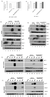
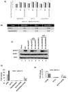
Type I IFN-induced antiviral state can inhibit the expression of MYXV early and late proteins in a species-specific manner; (A) Rabbit RK13 and (C) Mouse MEF PKR-/- cells were treated with hIFNβ and mIFNβ containing media for 18 h and infected with vMyx-FLuc (WT-MYXV) or vMyxM029KO-FLuc viruses for the indicated time points. Cells were then processed for luciferase assay. Percent reduction in FLuc expression was calculated from the samples not pretreated with IFNs. The assay was performed in triplicate; (B) Rabbit RK13 cells were treated with mock or hIFNβ containing media for 18 h and infected with WT-MYXV or vMyxM029KO viruses and cells were collected at the indicated time points for Western blot analysis; (D) Mouse MEFs and MEF PKR-/- cells were treated with mock or mIFNβ containing media for 18 h and infected with WT-MYXV or vMyxM029KO viruses and cells were collected at he indicated time points for Western blot analysis; (E) Human HeLa control and (F) HeLa PKR-/- cells were treated with mock (lanes 1-7) or hIFNβ (lanes 8-14) containing media for 18 h and infected with WT-MYXV or vMyxM029KO viruses and cells were collected at the indicated time points for Western blot analysis.
Figure 8
Figure 8
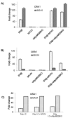
MYXV infection does not alter levels of ISGs in IFN pre-treated cells originating from different species; (A) Human HeLa PKR-/- and (B) Mouse MEF PKR-/- cells were treated with hIFNβ and mIFNβ respectively for 18h and infected with either WT-MYXV or vMyxM029KO viruses for another 24 h. Total RNA was isolated from the harvested cells and subjected to reverse transcription and real-time polymerase chain reaction (qPCR) to monitor the expression of ISGs. Fold changes are based on GAPDH as control; (C) Rabbit RK13 cells were transfected with poly I:C for 18 h and infected with either WT-MYXV or vMyxM029KO viruses for 24 h and total RNA was isolated from the harvested cells and subjected to reverse transcription and qPCR to monitor the expression of ISGs. Fold changes are based on GAPDH as control.
Figure 9
Figure 9
MYXV infection is able to inhibit post-infection type I IFN signaling even in the absence of M029; (A) MEF PKR-/-, RK13 and HeLa PKR-/- cells were infected with WT-MYXV and vMyxM029KO viruses alone for 1 h (adsorption) and 6 h, after which media containing mIFNβ or hIFNβ (500 U/mL) were added in one of the sample and harvested after 48 h for virus titration; (B) Table showing log10 changes in virus titer after different type I IFN treatment compare to the untreated samples at 48 h in MEF PKR-/-, RK13 and HeLa PKR-/- cells; (C) HeLa cells were left untreated, treated with hIFNβ for 30 mn, infected with vMyxGFP and vMyxM029KO viruses alone for 1 h and 6 h or treated with hIFNβ for 30 mn post-infection. The membranes were first probed with anti-pSTAT1 antibody, stripped and probed for STAT1 and again stripped and probed for actin (loading control); (D) HeLa PKR-/- cells were treated with hIFNβ for 18h after mock or infection with vMyxGFP or vMyxM029KO viruses for 6 h and total RNA was isolated from the cells and subjected to reverse transcription and qPCR to monitor the expression of Mx1 and ISG15. Fold changes are based on GAPDH as control; (E) MEF PKR-/- cells were treated with mIFNβ for 18 h after mock or infection with vMyxGFP for 6 h and total RNA was isolated from the cells and subjected to reverse transcription and qPCR to monitor the expression of Mx1 and ISG15. ** p < 0.01 *** p < 0.001. Fold changes are based on GAPDH as control.

Similar articles

Cited by

References

    1. Hoffmann H.H., Schneider W.M., Rice C.M. Interferons and viruses: An evolutionary arms race of molecular interactions. Trends Immunol. 2015;36:124–138. doi: 10.1016/j.it.2015.01.004. - DOI - PMC - PubMed
    1. Stifter S.A., Feng C.G. Interfering with immunity: Detrimental role of type I IFNs during infection. J. Immunol. 2015;194:2455–2465. doi: 10.4049/jimmunol.1402794. - DOI - PubMed
    1. Kerr P.J., Ghedin E., DePasse J.V., Fitch A., Cattadori I.M., Hudson P.J., Tscharke D.C., Read A.F., Holmes E.C. Evolutionary history and attenuation of myxoma virus on two continents. PLoS Pathog. 2013;8:e1002950. doi: 10.1371/journal.ppat.1002950. - DOI - PMC - PubMed
    1. Kerr P.J., Liu J., Cattadori I., Ghedin E., Read A.F., Holmes E.C. Myxoma virus and the Leporipoxviruses: An evolutionary paradigm. Viruses. 2015;7:1020–1061. doi: 10.3390/v7031020. - DOI - PMC - PubMed
    1. Werden S.J., Rahman M.M., McFadden G. Poxvirus host range genes. Adv. Virus Res. 2008;71:135–171. - PubMed

Publication types

LinkOut - more resources