. 2017 Apr;24(4):683-693.
doi: 10.1038/cdd.2017.1. Epub 2017 Feb 3.

Autophagy induced by DAMPs facilitates the inflammation response in lungs undergoing ischemia-reperfusion injury through promoting TRAF6 ubiquitination

Affiliations

Autophagy induced by DAMPs facilitates the inflammation response in lungs undergoing ischemia-reperfusion injury through promoting TRAF6 ubiquitination

Xingguang Liu et al. Cell Death Differ. 2017 Apr.

Abstract

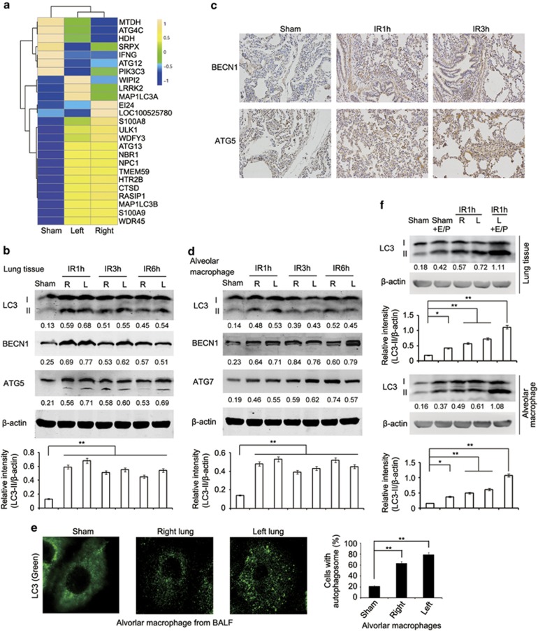
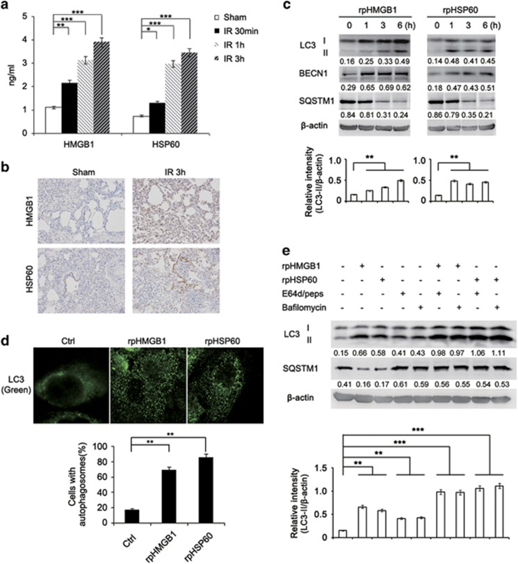
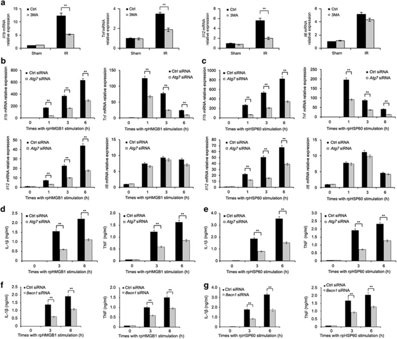
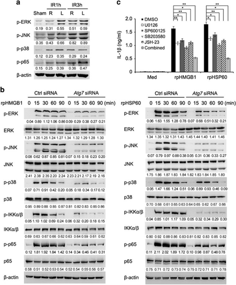
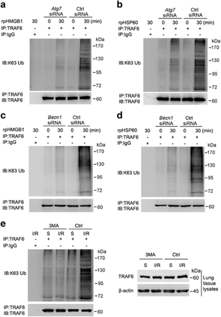
Lung ischemia-reperfusion (I/R) injury remains one of the most common complications after various cardiopulmonary surgeries. The inflammation response triggered by the released damage-associated molecular patterns (DAMPs) aggravates lung tissue damage. However, little is known about the role of autophagy in the pathogenesis of lung I/R injury. Here, we report that a variety of inflammation-related and autophagy-associated genes are rapidly upregulated, which facilitate the inflammation response in a minipig lung I/R injury model. Left lung I/R injury triggered inflammatory cytokine production and activated the autophagy flux as evidenced in crude lung tissues and alveolar macrophages. This was associated with the release of DAMPs, such as high mobility group protein B1 (HMGB1) and heat shock protein 60 (HSP60). Indeed, treatment with recombinant HMGB1 or HSP60 induced autophagy in alveolar macrophages, whereas autophagy inhibition by knockdown of ATG7 or BECN1 markedly reduced DAMP-triggered production of inflammatory cytokines including IL-1β, TNF and IL12 in alveolar macrophages. This appeared to be because of decreased activation of MAPK and NF-κB signaling. Furthermore, knockdown of ATG7 or BECN1 inhibited Lys63 (K63)-linked ubiquitination of TNF receptor-associated factor 6 (TRAF6) in DAMP-treated alveolar macrophages. Consistently, treatment with 3-MA inhibited K63-linked ubiquitination of TRAF6 in I/R-injured lung tissues in vivo. Collectively, these results indicate that autophagy triggered by DAMPs during lung I/R injury amplifies the inflammatory response through enhancing K63-linked ubiquitination of TRAF6 and activation of the downstream MAPK and NF-κB signaling.


Conflict of interest statement

The authors declare no conflict of interest.

Figures

Figure 1
Figure 1
Lung I/R injury triggers the inflammation response in lung tissues of minipigs. Different groups of minipigs underwent 1 h left lung ischemia followed by reperfusion for 1 h (IR1h), 3 h (IR3h) or 6 h (IR6h), or only received sham operations (Sham). (a) Heat maps showing hierarchical clustering of upregulated expressed transcripts of inflammation-related genes in right or left lung tissues from the sham group or minipigs subjected to lung ischemia followed by reperfusion for 1 h. (b) qPCR analysis of Tnf, Il1b, Il12 and Il6 mRNA expression in right (R) or left (L) lung tissues from the sham group or minipigs undergoing lung I/R as indicated (n=3). (c) Immunohistochemical staining of IL-1β and TNF expression in left lung tissues from the sham group or minipigs undergoing lung I/R as indicated. (d) qPCR analysis of Tnf, Il1b, Il12 and Il6 mRNA expression in alveolar macrophages from the sham group or minipigs undergoing lung I/R as indicated (n=3). (e) Immunofluorescence staining of IL-1β, F4/80 (macrophage marker), Ly6G (neutrophil marker) and SP-C (alveolar epithelial cell marker) in left lung tissues from minipigs subjected to lung ischemia followed by reperfusion for 1 h. Original magnification, × 400. Data are representative (c and e), or mean±S.E.M. (b and d), of three individual experiments. *P<0.05, **P<0.01, ***P<0.001
Figure 2
Figure 2
Lung I/R injury induces autophagy in lung tissues of minipigs. (a) Heat maps showing hierarchical clustering of differentially expressed transcripts of autophagy related genes in right or left lung tissues from the sham group or minipigs subjected to lung ischemia followed by reperfusion for 1 h. (b) Immunoblotting analysis of LC3, BECN1, ATG5 and β-actin (as a loading control) in lysates of right or left lung tissue from the sham group or minipigs subjected to lung I/R as indicated. (c) Immunohistochemical staining of BECN1 and ATG5 in left lung tissue of the sham group or minipigs subjected to lung I/R as indicated. (d) Immunoblotting analysis of LC3, BECN1, ATG7 and β-actin (as a loading control) in lysates of alveolar macrophages from BALF of the sham group or minipigs subjected to lung I/R as indicated. (e) Immunofluorescence analysis of LC3 in alveolar macrophages from BALF of the sham group or minipigs subjected to lung ischemia followed by reperfusion for 1 h. Original magnification, × 630. Quantification of cells with autophagosomes was also shown. (f) Immunoblotting analysis of LC3 and β-actin (as a loading control) in lysates of lung tissues (upper) or alveolar macrophages (lower) from the sham group or minipigs subjected to 1 h left lung ischemia and perfusion with pulmonary protective solution containing E64d (15 μg/ml) and Pepstatin A (15 μg/ml) or DMSO followed by reperfusion for 1 h. The band densitometry was quantified using ImageJ software (National Institutes of Health, Bethesda, MD, USA). Values below lanes represent the relative intensities of the corresponding proteins (LC3-II, BECN1, ATG5 and ATG7) to β-actin in the same lane. The relative band intensities of LC3-II/β-actin were calculated from three independent experiments and shown as mean±S.E.M. Data are representative of three individual experiments (bf). *P<0.05, **P<0.01
Figure 3
Figure 3
DAMPs are involved in lung I/R injury-triggered autophagy in alveolar macrophages of minipigs. (a) ELISA of HMGB1 and HSP60 production in the BALF of the sham group or minipigs subjected to lung I/R as indicated (n=3). (b) Immunohistochemical staining of HMGB1 and HSP60 in left lung tissues of the sham group or minipigs subjected to lung I/R as indicated. (c) Immunoblotting analysis of LC3, BECN1, SQSTM1 and β-actin (as a loading control) in lysates of alveolar macrophages treated with rpHMGB1 (1 μg/ml) or rpHSP60 (1 μg/ml) for the indicated periods. (d) Immunofluorescence analysis of LC3 in alveolar macrophages stimulated with rpHMGB1 (1 μg/ml) or rpHSP60 (1 μg/ml) for 3 h. Original magnification, × 630. Quantification of cells with autophagosomes is also shown. (e) Immunoblotting analysis of LC3, SQSTM1 and β-actin (as a loading control) in lysates of alveolar macrophages with or without treatment with E64d (15 μg/ml), Pepstatin A (15 μg/ml) or Bafilomycin A1 (30 nM) before stimulation by rpHMGB1 (1 μg/ml) or rpHSP60 (1 μg/ml) for 3 h. Values below lanes represent the relative intensities of the corresponding proteins (LC3-II, BECN1 and SQSTM1) to β-actin in the same lane. The relative band intensities of LC3-II/β-actin were calculated from three independent experiments and shown as mean±S.E.M. Data are mean±S.E.M. (a) or representative (be), of three individual experiments. *P<0.05, **P<0.01, ***P<0.001
Figure 4
Figure 4
Treatment with 3-MA or ATG7 knockdown attenuates inflammatory cytokine production in lung tissues or alveolar macrophages respectively. (a) Minipigs were subjected to lung ischemia and perfusion with pulmonary protective solution containing 3-MA (5 mM) or DMSO followed by reperfusion for 1 h or received sham operations (Sham). The levels of Il1b, Tnf, Il12 and Il6 mRNA expression in left lung tissue were detected by qPCR analysis. (bg) Alveolar macrophages were transfected with the control siRNA, Atg7 siRNA or Becn1 siRNA. Sixty hour later, cells were treated with rpHMGB1 (1 μg/ml) (b, d and f) or rpHSP60 (1 μg/ml) (c, e and g) for the indicated periods. The levels of Il1b, Tnf, Il12 and Il6 mRNA expression were detected by qPCR analysis (b and c), or the levels of IL-1β and TNF in supernatants were measured by ELISA (dg). Data are mean±S.E.M. of three individual experiments. **P<0.01
Figure 5
Figure 5
Autophagy is involved in activation of MAPK and NF-κB signaling induced by DAMPs in alveolar macrophages of minipigs. (a) Immunoblotting analysis of phosphorylation (p−) levels of ERK, JNK, p38 and p65 in lysates of alveolar macrophages isolated from right or left lungs of minipigs in the sham group or those undergoing lung I/R as indicated. Values below lanes represent the relative intensities of the corresponding proteins to β-actin in the same lane. (b) Immunoblotting analysis of phosphorylation (p−) or total levels of ERK, JNK, p38, IKKα/β and p65 in lysates of alveolar macrophages transfected with the control siRNA or Atg7 siRNA followed by treatment with rpHMGB1 (1 μg/ml) (left) or rpHSP60 (1 μg/ml) (right) for the indicated periods. Values below lanes represent the relative intensities of the phosphorylated proteins to the total proteins or the total proteins to β-actin, respectively, in the same lane. (c) ELISA of IL-1β production in supernatants of alveolar macrophages with or without pretreatment with inhibitors of ERK (U0126), JNK (SP600125), p38 (SB203580) or p65 (JSH-23), or under the combined use of the above inhibitors for 30 min followed by stimulation with rpHMGB1 (1 μg/ml) or rpHSP60 (1 μg/ml) for 3 h. Data are representative (a and b), or mean±S.E.M. (c), of three individual experiments. *P<0.05, **P<0.01
Figure 6
Figure 6
Autophagy is involved in TRAF6 ubiquitination triggered by lung I/R injury. (ad) Alveolar macrophages were transfected with the control siRNA, Atg7 siRNA (a and b) or Becn1 siRNA (c and d). Sixty hours later, cells were treated with rpHMGB1 (1 μg/ml) (a and c) or rpHSP60 (1 μg/ml) (b and d) for 30 min or left untreated. (e) Minipigs underwent lung ischemia and perfusion with pulmonary protective solution containing 3-MA (5 mM) or DMSO followed by reperfusion for 30 min or received sham operations (Sham). Whole-cell lysates of alveolar macrophages (ad) or lysates of left lung tissues (e) were subjected to immunoprecipitation with anti-TRAF6 antibody followed by immunoblotting with anti-K63 ubiquitin and anti-TRAF6 antibodies, or immunoblotted with anti-TRAF6 and β-actin antibodies. S: sham group. Data are representative of three individual precipitation experiments

Similar articles

Cited by

References

    1. Eltzschig HK, Bratton DL, Colgan SP. Targeting hypoxia signalling for the treatment of ischaemic and inflammatory diseases. Nat Rev Drug Discov 2014; 13: 852–869. - PMC - PubMed
    1. van Golen RF, Reiniers MJ, Olthof PB, van Gulik TM, Heger M. Sterile inflammation in hepatic ischemia/reperfusion injury: present concepts and potential therapeutics. J Gastroenterol Hepatol 2013; 28: 394–400. - PubMed
    1. Tang D, Kang R, Coyne CB, Zeh HJ, Lotze MT. PAMPs and DAMPs: signal 0 s that spur autophagy and immunity. Immunol Rev 2012; 249: 158–175. - PMC - PubMed
    1. den Hengst WA, Gielis JF, Lin JY, Van Schil PE, De Windt LJ, Moens AL. Lung ischemia-reperfusion injury: a molecular and clinical view on a complex pathophysiological process. Am J Physiol Heart Circ Physiol 2010; 299: H1283–H1299. - PubMed
    1. Mizushima N, Levine B, Cuervo AM, Klionsky DJ. Autophagy fights disease through cellular self-digestion. Nature 2008; 451: 1069–1075. - PMC - PubMed

Publication types

MeSH terms