Review
. 2017 Feb;19(2):177-191.
doi: 10.1002/ejhf.696.

Myocardial fibrosis: biomedical research from bench to bedside

Affiliations
Review

Myocardial fibrosis: biomedical research from bench to bedside

Mariann Gyöngyösi et al. Eur J Heart Fail. 2017 Feb.

Abstract

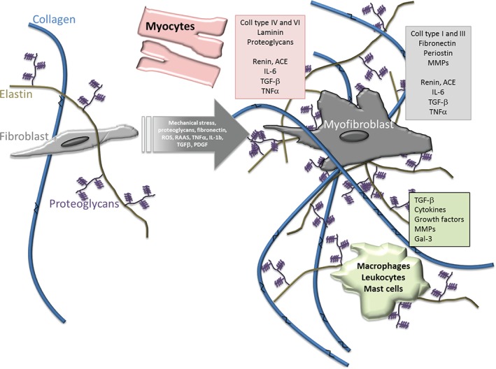
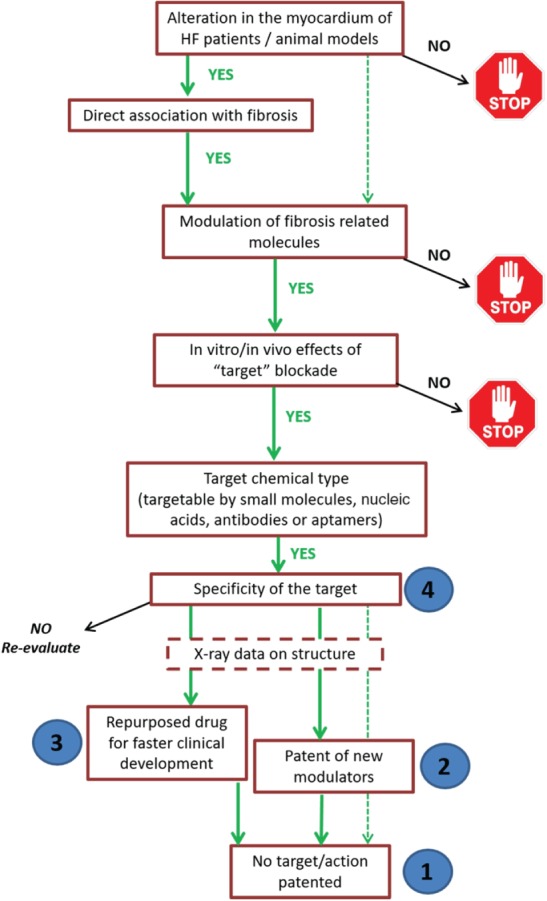
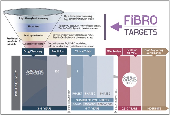
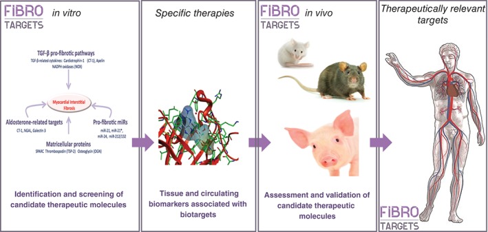
Myocardial fibrosis refers to a variety of quantitative and qualitative changes in the interstitial myocardial collagen network that occur in response to cardiac ischaemic insults, systemic diseases, drugs, or any other harmful stimulus affecting the circulatory system or the heart itself. Myocardial fibrosis alters the architecture of the myocardium, facilitating the development of cardiac dysfunction, also inducing arrhythmias, influencing the clinical course and outcome of heart failure patients. Focusing on myocardial fibrosis may potentially improve patient care through the targeted diagnosis and treatment of emerging fibrotic pathways. The European Commission funded the FIBROTARGETS consortium as a multinational academic and industrial consortium with the primary aim of performing a systematic and collaborative search of targets of myocardial fibrosis, and then translating these mechanisms into individualized diagnostic tools and specific therapeutic pharmacological options for heart failure. This review focuses on those methodological and technological aspects considered and developed by the consortium to facilitate the transfer of the new mechanistic knowledge on myocardial fibrosis into potential biomedical applications.

Keywords: Animal models; Biomarkers; Cardiac imaging; Myocardial fibrosis.


Figures

Figure 1
Figure 1
Schematic representation of biochemical and cellular mechanisms of cardiac fibrosis. Under physiological conditions (left), fibroblasts secrete extracellular procollagen chains into the interstitium that assemble into fibrils and are cross‐linked by lysyl oxidase. Several cell types are implicated in fibrotic remodelling of the heart either directly by producing matrix proteins (fibroblasts), or indirectly by secreting fibrogenic mediators (macrophages, mast cells, lymphocytes, cardiomyocytes, and vascular cells). Under pathological conditions (right), alterations in the matrix environment, induction and release of growth factors and cytokines, and increase of mechanical stress dynamically modulate fibroblast transdifferentiation into myofibroblasts. Higher collagen cross‐linking results in increased myocardial tensile strength. Resistance to degradation by matrix metalloproteinases (MMPs) increases cross‐linked collagen, which favours matrisome expansion. Pink, grey, and green boxes list part of the secretome of mycocytes, myofibroblasts, and macrophages/leucocytes/mast cells, respectively, that trigger and maintain fibrosis. Gal‐3, galectin‐3; IL, interleukin; PDGF, platelet‐derived growth factor; RAAS, renin–angiotensin–aldosterone system; ROS, reactive oxygen species; TGF, transforming growth factor; TNF, tumour necrosis factor.
Figure 2
Figure 2
Representative native and T1 cardiac magnetic resonance imaging (cMRI) of diffuse myocardial fibrosis. (A) Diffuse myocardial fibrosis on the short‐axis view of the cMRI image, with the circumference of the anteroseptal myocardial area (region of interest). (B) cMRI T1 map of a patient with moderate aortic stenosis and moderate diffuse myocardial fibrosis. (C) cMRI T1 map of another patient with severe aortic stenosis and severe diffuse fibrosis of the left ventricle. Reproduced with permission from the Radiological Society of North America from Lee et al. 76
Figure 3
Figure 3
Algorithm for selection of new antifibrotic factors to be further tested as potential therapeutic targets. In order to prioritize the potential antifibrotic targets currently under study in the FIBROTARGETS consortium, and select those to be evaluated in depth from a therapeutic point of view, a number of aspects will be considered in a step‐by‐step process. Targets need to fulfil the stated criteria, otherwise they will be discarded (stop signs). Numbers in blue circles indicate the prioritization of potential therapeutic agents according to their properties. HF, heart failure.
Figure 4
Figure 4
Drug development pipeline highlighting the phases developed by the FIBROTARGETS consortium (modified from Phrma.com). The activities developed by the consortium cover the first steps of the drug discovery strategy; high throughput screening (HTS), hit to lead phase, and lead optimization. By the end of the project, we aim to have identified a set of promising candidates for further evaluation. ADME, absorption, distribution, metabolism, and excretion; FDA, Food and Drug Administration; IC50, half‐maximal inhibitory concentration; HCS, high content screening; IND, investigational new drug; MFG, manufacturing; NDA, new drug application; PD, pharmacodynamics; PK, pharmacokinetics; POC, proof of concept.
Figure 5
Figure 5
Schematic workflow and aims of FIBROTARGETS.

References

    1. Weber KT, Sun Y, Bhattacharya SK, Ahokas RA, Gerling IC. Myofibroblast‐mediated mechanisms of pathological remodelling of the heart. Nat Rev Cardiol 2013;10:15–26. - PubMed
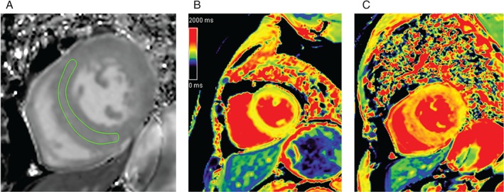
    1. Jellis C, Martin J, Narula J, Marwick TH. Assessment of nonischemic myocardial fibrosis. J Am Coll Cardiol 2010;56:89–97. - PubMed
    1. Kong P, Christia P, Frangogiannis N. The pathogenesis of cardiac fibrosis. Cell Mol Life Sci 2014;71:549–574. - PMC - PubMed
    1. Li AH, Liu PP, Villarreal FJ, Garcia RA. Dynamic changes in myocardial matrix and relevance to disease: translational perspectives. Circ Res 2014;114:916–927. - PubMed
    1. Weber KT, Pick R, Jalil JE, Janicki JS, Carroll EP. Patterns of myocardial fibrosis. J Mol Cell Cardiol 1989;21:121–131. - PubMed