. 2017 Feb 3;12(2):e0171510.
doi: 10.1371/journal.pone.0171510. eCollection 2017.

RB inactivation in keratin 18 positive thymic epithelial cells promotes non-cell autonomous T cell hyperproliferation in genetically engineered mice

Affiliations

RB inactivation in keratin 18 positive thymic epithelial cells promotes non-cell autonomous T cell hyperproliferation in genetically engineered mice

Yurong Song et al. PLoS One. .

Abstract

Thymic epithelial cells (TEC), as part of thymic stroma, provide essential growth factors/cytokines and self-antigens to support T cell development and selection. Deletion of Rb family proteins in adult thymic stroma leads to T cell hyperplasia in vivo. To determine whether deletion of Rb specifically in keratin (K) 18 positive TEC was sufficient for thymocyte hyperplasia, we conditionally inactivated Rb and its family members p107 and p130 in K18+ TEC in genetically engineered mice (TgK18GT121; K18 mice). We found that thymocyte hyperproliferation was induced in mice with Rb inactivation in K18+ TEC, while normal T cell development was maintained; suggesting that inactivation of Rb specifically in K18+ TEC was sufficient and responsible for the phenotype. Transplantation of wild type bone marrow cells into mice with Rb inactivation in K18+ TEC resulted in donor T lymphocyte hyperplasia confirming the non-cell autonomous requirement for Rb proteins in K18+ TEC in regulating T cell proliferation. Our data suggests that thymic epithelial cells play an important role in regulating lymphoid proliferation and thymus size.


Conflict of interest statement

The authors have declared that no competing interests exist. KK, DCH and JK are government contract employees of Leidos Biomedical Research, Inc., who work in the Frederick National Laboratory for Cancer Research (FNLCR). This does not alter our adherence to PLOS ONE policies on sharing data and materials.

Figures
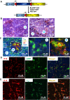
Fig 1
Fig 1. Transgene expression by immunostaining in TgK18GT121 (K18) or induced-K18 mice.
(A) Transgene cassette consisting of floxed eGFP stop cassette upstream of truncated SV40 large T antigen (first 121 amino acid; T121) was inserted into the 1st exon of K18 gene on a bacterial artificial chromosome (BAC). Transgene eGFP was driven by K18 regulation. Once K18 mice were crossed to a transgenic mice expressing Cre recombinase, T121 was expressed directly under K18 regulation. (B) Representative images of K18 IHC staining in cortex (C) and medulla (M) of WT thymus. Inserts are higher magnification of the images. (C) Representative immunofluorescence images of T121 (green), K18 (yellow), K5 (red), and DAPI (blue) in cortex (C) and medulla (M) delineated with a white dotted line, in induced-K18;Cre thymus. Middle and right images are higher magnification of areas in white and red boxes of left image, respectively. Right image: * Cell is positive for T121, K18, and K5, and # positive for K5 only. (D) Representative images of K18 (red) and eGFP (green) immunostaining in thymic cortex and medulla (data not shown) of uninduced-K18 mice (Cre negative). (E) Representative images of K18 (red) and T121 (green) immunostaining in thymic cortex and medulla (data not shown) of induced-K18 mice (K18;β-actin Cre).
Fig 2
Fig 2. Rb-TS inactivation in K18+ TEC led to decreased survival and thymic hyperplasia.
(A) Kaplan-Meier survival curve of K18;β-actin Cre (n = 74), K18;R26CreER (n = 27), and K18;PbCre4 (n = 45) mice with median survival of 94, 41, and 231 days, respectively. Uninduced -K18 mice (n = 8) did not develop any gross abnormalities. (B) Gross phenotype of thymuses in WT and K18;β-actin Cre mice. (C) Representative images of H.E. stained thymus sections in WT, K18;R26CreER mice. C: cortex; M: medulla. (D) Representative low magnification images of H.E. stained thymuses in WT and K18;β-actin Cre mice. C: cortex; M: medulla.
Fig 3
Fig 3. T cell populations are not altered by Rb-TS inactivation in K18+ TEC.
(A) Total thymic cellularity (left); thymus weight (middle) and spleen weight (right) in grams (g) in WT (n = 5) and K18;Cre (n = 10) mice. Data are presented as mean ± SEM. p<0.05 is considered statistically significant. (B) Representative FACS plots of CD4 and CD8 staining in WT, K18;R26CreER, K18;β-actin Cre, and K18;PbCre4 thymuses. (C) Percentage of CD4+, CD4+CD8+, CD8+, and CD4-CD8- T cell subpopulations in thymuses of WT (1, n = 5), K18;R26CreER (2, n = 8), K18;β-actin Cre (3, n = 3), and K18;PbCre4 (4, n = 13) (top, no statistically significant difference among cohorts), and T cell subpopulations in spleens (bottom) of WT (n = 4) and K18;Cre (n = 17) by FACS analysis. p<0.05 is considered statistically significant.
Fig 4
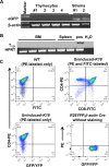
Fig 4. Transgene is not expressed in thymocytes.
(A) eGFP mRNA levels in thymocytes and thymic stroma of uninduced-K18 mice by RT-PCR. Thymocytes was released by gently applying pressure on thymus between two frosted glass slides, and then FACS-sorted for CD45+ cells. CD45- cells were used as thymic stroma and served as positive control. RNA was extracted from both CD45+ thymocytes and CD45- thymic stroma for RT-PCR. β-actin was used as loading control. (B) T121 mRNA levels in bone marrow (BM) and spleen of K18;PbCre4 mice by RT-PCR. Bone marrow cell suspension was obtained by flushing the femurs with PBS. CD45- thymic stroma obtained for Fig 4A was used as positive control (pos), and HPRT as loading control. (C) Representative FACS plot to assess whether eGFP was expressed in thymocytes of uninduced-K18 mice. Thymocytes were stained with only CD4-PE antibody in WT and uninduced-K18 mice (left panel), or with both CD4-PE and CD8-FITC antibodies in uninduced-K18 mice (top right). YFP in thymocytes of R26YFP;β-actin Cre mice without any antibody staining was readily detected and used as a positive control (bottom right).
Fig 5
Fig 5. Rb-TS inactivation in K18+ TEC extrinsically regulates thymic hyperplasia.
(A) Bone marrow cells including hematopoietic stem and progenitor cells and mature hematopoietic cells (erythroid, myeloid, B cells), as well as very few stromal cells of mesenchymal and endothelial lineages were obtained from Ly5.1+ wildtype (WT, n = 7) and K18;PbCre4 (n = 9) mice, and—transplanted into 3-month old Ly5.2+ lethally irradiated WT recipients. There is no statistical difference between WT and K18;PbCre4 donor group. (a) Weight of thymus and spleen in the reconstituted recipients with either WT or K18;PbCre4 bone marrow cells as donor. (b) Percentage of thymocytes derived from WT or K18;PbCre4 donors. DP: double positive (CD4+CD8+), DN: double negative (CD4-CD8-). Data is presented as mean ± SEM. (B) Bone marrow cells obtained from Ly5.2+ WT mice were transplanted into 3-month old Ly5.1+ irradiated K18;PbCre4 recipients (K18 Rec, n = 5; three recipients were found dead without thymus enlargement at 2–3 weeks post transplantation) or WT (WT Rec, n = 11) recipients. (a) Weight of thymus and spleen of the reconstituted recipients in grams (g), and mo: months post transplantation. * p < 0.001 (4mo K18 Rec vs. WT Rec). (b) Percentage of T cell subpopulations in WT Rec and K18 Rec transplanted with WT bone marrow cells by FACS analysis. DP: double positive (CD4+CD8+); DN: double negative (CD4-CD8-).

Similar articles

Cited by

References

    1. Boyd RL, Tucek CL, Godfrey DI, Izon DJ, Wilson TJ, Davidson NJ, et al. The thymic microenvironment. Immunol Today. 1993;14(9):445–59. 10.1016/0167-5699(93)90248-J - DOI - PubMed
    1. Gray DH, Ueno T, Chidgey AP, Malin M, Goldberg GL, Takahama Y, et al. Controlling the thymic microenvironment. Curr Opin Immunol. 2005;17(2):137–43. 10.1016/j.coi.2005.02.001 - DOI - PubMed
    1. Chu PG, Weiss LM. Keratin expression in human tissues and neoplasms. Histopathology. 2002;40(5):403–39. Epub 2002/05/16. - PubMed
    1. Omary MB, Ku NO, Strnad P, Hanada S. Toward unraveling the complexity of simple epithelial keratins in human disease. J Clin Invest. 2009;119(7):1794–805. Epub 2009/07/10. PubMed Central PMCID: PMC2701867. 10.1172/JCI37762 - DOI - PMC - PubMed
    1. Moll R, Divo M, Langbein L. The human keratins: biology and pathology. Histochem Cell Biol. 2008;129(6):705–33. Epub 2008/05/08. PubMed Central PMCID: PMC2386534. 10.1007/s00418-008-0435-6 - DOI - PMC - PubMed

LinkOut - more resources