. 2017 Apr 20;45(7):3844-3859.
doi: 10.1093/nar/gkx065.

WRN is recruited to damaged telomeres via its RQC domain and tankyrase1-mediated poly-ADP-ribosylation of TRF1

Affiliations

WRN is recruited to damaged telomeres via its RQC domain and tankyrase1-mediated poly-ADP-ribosylation of TRF1

Luxi Sun et al. Nucleic Acids Res. .

Abstract

Werner syndrome (WS) is a progeroid-like syndrome caused by WRN gene mutations. WS cells exhibit shorter telomere length compared to normal cells, but it is not fully understood how WRN deficiency leads directly to telomere dysfunction. By generating localized telomere-specific DNA damage in a real-time fashion and a dose-dependent manner, we found that the damage response of WRN at telomeres relies on its RQC domain, which is different from the canonical damage response at genomic sites via its HRDC domain. We showed that in addition to steady state telomere erosion, WRN depleted cells are also sensitive to telomeric damage. WRN responds to site-specific telomeric damage via its RQC domain, interacting at Lysine 1016 and Phenylalanine1037 with the N-terminal acidic domain of the telomere shelterin protein TRF1 and demonstrating a novel mechanism for WRN's role in telomere protection. We also found that tankyrase1-mediated poly-ADP-ribosylation of TRF1 is important for both the interaction between WRN and TRF1 and the damage recruitment of WRN to telomeres. Mutations of potential tankyrase1 ADP-ribosylation sites within the RGCADG motif of TRF1 strongly diminish the interaction with WRN and the damage response of WRN only at telomeres. Taken together, our results reveal a novel mechanism as to how WRN protects telomere integrity from damage and telomere erosion.


Figures

Figure 1.
Figure 1.
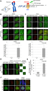
WRN is recruited to sites of oxidative DNA damage at telomeres. (A) Overview of the Damage Targeted at Telomeres method to induce ROS damage specifically at sites of telomeres. KillerRed (KR) is fused with TRF1 to bind to telomeric DNA repeats. Exposure of cells to visible light will activate KR to induce oxidative DNA damage at telomeres in living cells. (B and C) GFP-WRN and KR-TRF1, or DsR-TRF1 or RFP-TRF1, were co-transfected into U2OS cells (B) and HeLa cells (C). The single cell nucleus was scanned by a 559 nm laser; recruitment of WRN to KR-TRF1 damage sites in both U2OS (B) and HeLa cells (C) 3 min after activation is shown (upper panel). Quantification of the damage response of GFP-WRN at sites of KR-TRF1 (lower panel). The fold increase of GFP-WRN (mean intensity of WRN at KR-TRF1/mean intensity of GFP-WRN distant from KR-TRF1 in the nucleus) is shown in the U2OS cell line (B) and HeLa cell line (C). Data are represented as mean ± SEM, **P < 0.005. (D) GFP-WRN and KR-TRF1 were co-transfected into U2OS cells and one KR-TRF1 spot (indicated by a yellow square) was scanned by the 559 nm laser to activate KR; recruitment of WRN to the single KR-TRF1 spot is shown 3 min after activation. GFP-WRN at an inactivated KR-TRF1 spot is shown in the blue square. (E and F) Damage response of endogenous WRN at sites of telomeres is shown. U2OS cells synchronized at G0/G1 phase expressing either DsR-TRF1 or KR-TRF1 were exposed to a 15 W SYLVANIA cool white fluorescent bulb for 20 min and then immunostained with WRN antibody. Percentage of colocalization of WRN and DsR-TRF1 or KR-TRF1 (>5 colocalization spots were defined as positive) is shown. A total of 150 cells in total were counted in three independent experiments, data are represented as mean ± SEM.
Figure 2.
Figure 2.
The RQC domain of WRN responds to oxidative damage at telomeres but not at genome sites. (A) Schematic representation of the DDR of a full length (FL) GFP-tagged WRN and truncated or mutated WRN at telomeres and genome sites. (+) means a positive damage response and (−) means no damage response. (B) Three min after 559 nm laser light activation of KR, recruitment of GFP-tagged truncated or mutated WRN proteins to DsRed or KR-TRF1-induced damage at telomeres is shown in U2OS cells. The yellow rectangle indicates an enlarged area showing the colocalization of WRN at the sites of KR-TRF1. (C) Upper panel: recruitment of RQC domain of WRN at telomeres after KR-TRF1 activation in HeLa cells. Lower panel: Scheme of Damage Targeted at Genomic sites method to induce ROS damage specifically at one genome locus of a chromosome is shown. The tetracycline repressor (tetR) fused to KR (tetR-KR) binds a tetracycline response element (TRE) cassette in the defined genome site in U2OS cells. (D) Recruitment of GFP-tagged WRN FL and the RQC domain to 405 nm laser induced damage before (left) and 3 min after (right) 405 nm laser irradiation for 500 ms. The DDR of WRN FL but not the RQC domain to tetR-KR (D) or 405 nm laser (E) is shown with yellow arrowheads. (E) Quantification of accumulation kinetics of GFP-tagged WRN FL and the RQC domain at sites of telomeric damage by the fold increase of the relative intensity after 559 nm laser light irradiation. (F) Dissociation kinetics of GFP-tagged WRN FL and the RQC domain at sites of telomeric damage at 10 min, 24 and 48 h recovery time after 20 min cool white fluorescent bulb light activation of KR. The percentages of co-localization of WRN FL or the RQC domain with KR-TRF1 are shown in the graph. A total of 150 cells in total were counted in three independent experiments; data are represented as mean ± SEM.
Figure 3.
Figure 3.
WRN and BER factors are recruited to oxidative damage at telomeres independently. (A) Recruitment of WRN FL and RQC was observed in U2OS cells after siTRF2 treatment. Images before or 3 min after 559 nm laser light exposure or without laser light exposure of KR-TRF1 are shown. Yellow rectangles indicate the enlarged area. (B) Recruitment of WRN was observed 3 min after 559 nm laser light exposure of KR-TRF1 in WT and PARP1−/− and Polβ−/− MEF cells. (C) Dissociation of WRN from telomeres after damage induction was delayed by knocking down PARP1 or FEN1. Cells were exposed to light for 20 min and then placed in the dark for 48 h before fixation. The percentages of co-localization of WRN FL or RQC with KR-TRF1 are shown in the graph. A total of 150 cells in total were counted in three independent experiments; data are represented as mean ± SEM.
Figure 4.
Figure 4.
WRN interacts with TRF1 after induction of oxidative damage at telomeres via the RQC domain of WRN and the N-terminal acidic domain of TRF1. (A) Schematic representation of the experimental procedure for IP. (B) WRN interacts with TRF1 after KR-TRF1 activation. KR-TRF1 stably expressing Flp-in T-REx 293 cells were transfected with GFP-WRN. Tetracycline was added to induce KR-TRF1 expression 48 h before light illumination. Cells were treated with or without 20 min cool white fluorescent bulb light exposure to activate KR and incubated in the dark for 10 min (all the same treatments in 4C-4F before IP). Cell lysates were immunoprecipitated with or without α-TRF1. The precipitates and 3% of the lysate (input) were immunoblotted with α-GFP and α-TRF1. (C) GFP-tagged WRN FL and RQC were transfected into KR-TRF1 stably expressing Flp-in T-REx 293 cells, respectively. Cell lysates were immunoprecipitated with α-TRF1 and immunoblotted with α-GFP and α-TRF1. Arrowheads indicate the bands of GFP-tagged WRN truncations. (D and E) Schematic representation of domains of TRF1 and truncations of Myc-tagged TRF1 are shown. Mapping the interaction domain of TRF1 with WRN and WRN RQC. KR-TRF1 stably expressing Flp-in T-REx 293 cells were cotransfected with Myc-tagged TRF1 deletions shown in 4C and GFP-WRN (D) or GFP-WRN-RQC (E). Cell lysates were immunoprecipitated with α-Myc and immunoblotted with α-myc and α-GFP. (F) GFP-tagged WRN RQC, RQC K1016A and Phe1037A were transfected into FLAG-KR-TRF1 stably expressing Flp-in T-REx 293 cells, respectively. Cell lysates were immunoprecipitated with α-FLAG and immunoblotted with α-GFP and α-FLAG. Arrowheads indicate the bands of GFP-tagged WRN mutants. (G) Upper: before and 3 min after 559 nm laser light activation of KR, recruitment of GFP-tagged WRN RQC (K1016A and Phe1037A) proteins to KR-TRF1-induced damage at telomeres is shown in U2OS cells. Middle: Quantification of accumulation of GFP-tagged WRN-RQC, RQC K1016A and Phe1037A at sites of telomeric damage by the fold increase of the relative intensity after 559 nm laser light irradiation (n = 10). (H) Schematic representation of the interaction between WRN and TRF1 mediated by the RQC domain of WRN and the N-terminal acidic domain of TRF1.
Figure 5.
Figure 5.
TNKS1 mediated poly (ADP-ribosyl) ation of TRF1 after damage is necessary for the recruitment of WRN to sites of telomeric damage. (A) TNKS1 is recruited to sites of KR-TRF1 induced damage after light activation. KR-TRF1 transfected U2OS cells (left) were stained with α-TNKS1 after light exposure for 20 min. The yellow rectangle indicates the enlarged area. (B) TNKS1-mediated poly (ADP-ribosyl) ation of TRF1 is induced by telomeric oxidative damage. KR-TRF1 stably expressing HeLa cells were treated with or without siTNKS1 or 92 nM G007-LK for 24 h, or 4 μM PJ34 for 30 min. Cell lysates were collected with the lysis buffer containing 960 μM of the PARG inhibitor, ADP-HPD dihydrate ammonium salt, immediately after light exposure and immunoprecipitated with α-TRF1. The precipitates and 3% of the lysate (input) were immunoblotted with α-pAR and α-TRF1. (C) Recruitment of WRN to oxidative damage at telomeres is prevented by siTNKS1 or PARP inhibitors. Recruitment of GFP-WRN to KR-TRF1 induced damage at telomeres 3 min after 559 nm laser bleaching with treatment of Olaparib, PJ34, G007-LK or siTNKS1 in U2OS cells (left). Quantification of the damage response of GFP-WRN at sites of KR-TRF1 is shown (right). Data are represented as mean ± SEM of 10 cells. **P < 0.005.
Figure 6.
Figure 6.
The interaction between WRN or WRN-RQC and TRF1 is dependent on TNKS1-mediated PARylation. GFP-WRN (A and C) or GFP-WRN-RQC (B, D and E) and Myc-TRF1-N-A were co-transfected into KR-TRF1 stably expressing Flp-in T-REx 293 cells. After KR-TRF1 expression was induced by tetracycline, cells were treated with or without either PJ34 or G007-LK. Cells were treated with 20 min light exposure and recovered in the dark for 10 min. Cell lysates were immunoprecipitated with α-Myc. The precipitates and 3% of the lysate (input) were immunoblotted with α-GFP antibody and α-Myc antibody. Arrowheads indicate the bands of immunoprecipitated WRN FL or RQC. (F) TBM mutant overexpression diminished the interaction with and recruitment of WRN after telomeric damage. Left: 293 cells were transfected with FLAG-TRF1 or TBM and GFP-RQC. After damage induction, cell lysates were immunoprecipitated with α-GFP and immunoblotted with α-FLAG. Middle: schematic illustration of the TBM mutant is shown. Right: U2OS cells were co-transfected with GFP-WRN, KR-TRF2 and FLAG-TRF1 WT or TBM. Cells were treated with 20 min light exposure and recovered in the dark for 10 min. The percentages of co-localization of WRN FL or RQC with KR-TRF1 are shown in the graph. A total of 150 cells in total were counted in three independent experiments; data are represented as mean ± SEM.
Figure 7.
Figure 7.
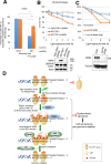
Suppression of WRN causes delayed repair and increased cell death after oxidative damage at telomeres. (A) Quantification of the percentage of cells showing co-localization of KR-TRF1 and γH2AX with or without siWRN at the indicated recovery time point after 20 min of 15 W SYLVANIA cool white fluorescent bulb light activation of KR. Data are represented as mean ± SEM of three independent experiments with counting 150 cells/time. **P < 0.005 (B and C) Clonogenic survival assay of HeLa cells with stably expressed KR-TRF1 or DsR-TRF1 and treated with or without siWRN (B), siUTR-WRN (C) with or without expression of WRN-RQC. Cells were exposed to cool white fluorescent light for the indicated time. Data are represented as mean ± SEM of three independent experiments. Western blot analysis of WRN knockdown and DsR-TRF1 and KR-TRF1 expression in HeLa cells is shown. (D) A model of recruitment of WRN to oxidative DNA damage at telomeres via interaction with TRF1 upon TNKS1-mediated PARylation. Oxidative DNA damage at telomeres induced a TRF1 conformational change to expose its N-terminal acidic domain. The N-terminal acidic domain of TRF1 will be targeted for PARylation by TNKS1 and then WRN is recruited to sites of DNA damage mediated by interaction between the RQC domain of WRN and N-terminal PARylation of the acidic domain of TRF1. BER factors are recruited independently from WRN. WRN protein dissociates from the sites of damage after repair completion. The function of WRN at telomeres protects genome stability in the face of oxidative DNA damage.

Similar articles

Cited by

References

    1. Cooke M.S., Evans M.D., Dizdaroglu M., Lunec J.. Oxidative DNA damage: mechanisms, mutation, and disease. FASEB J. 2003; 17:1195–1214. - PubMed
    1. Hegde M.L., Mantha A.K., Hazra T.K., Bhakat K.K., Mitra S., Szczesny B.. Oxidative genome damage and its repair: implications in aging and neurodegenerative diseases. Mech. Ageing Dev. 2012; 133:157–168. - PMC - PubMed
    1. Wang Z., Rhee D.B., Lu J., Bohr C.T., Zhou F., Vallabhaneni H., de Souza-Pinto N.C., Liu Y.. Characterization of oxidative guanine damage and repair in mammalian telomeres. PLoS Genet. 2010; 6:e1000951. - PMC - PubMed
    1. Sahin E., Depinho R.A.. Linking functional decline of telomeres, mitochondria and stem cells during ageing. Nature. 2010; 464:520–528. - PMC - PubMed
    1. Richter T., von Zglinicki T.. A continuous correlation between oxidative stress and telomere shortening in fibroblasts. Exp. Gerontol. 2007; 42:1039–1042. - PubMed

Publication types

LinkOut - more resources