. 2017 Feb 3;6(2):e004161.
doi: 10.1161/JAHA.116.004161.

Tissue Transglutaminase Modulates Vascular Stiffness and Function Through Crosslinking-Dependent and Crosslinking-Independent Functions

Affiliations

Tissue Transglutaminase Modulates Vascular Stiffness and Function Through Crosslinking-Dependent and Crosslinking-Independent Functions

Jochen Steppan et al. J Am Heart Assoc. .

Abstract

Background: The structural elements of the vascular wall, namely, extracellular matrix and smooth muscle cells (SMCs), contribute to the overall stiffness of the vessel. In this study, we examined the crosslinking-dependent and crosslinking-independent roles of tissue transglutaminase (TG2) in vascular function and stiffness.

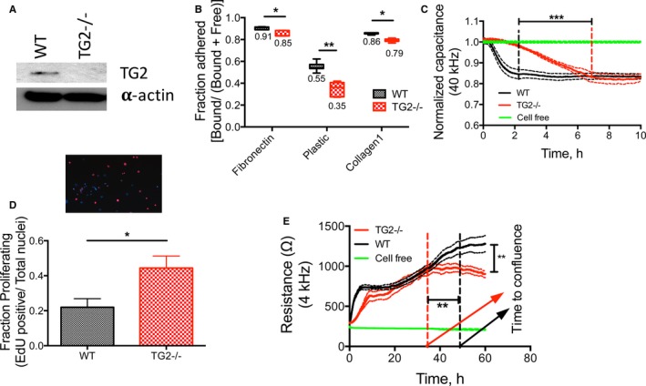
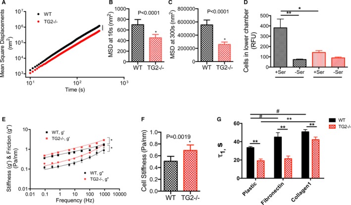
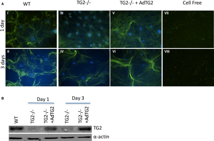
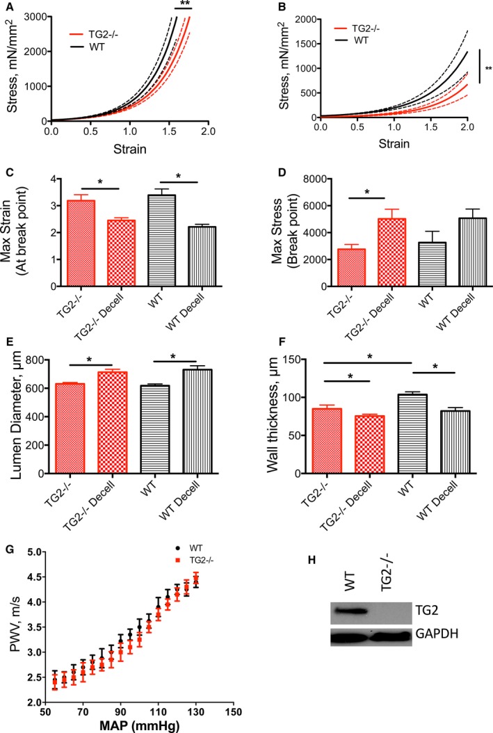
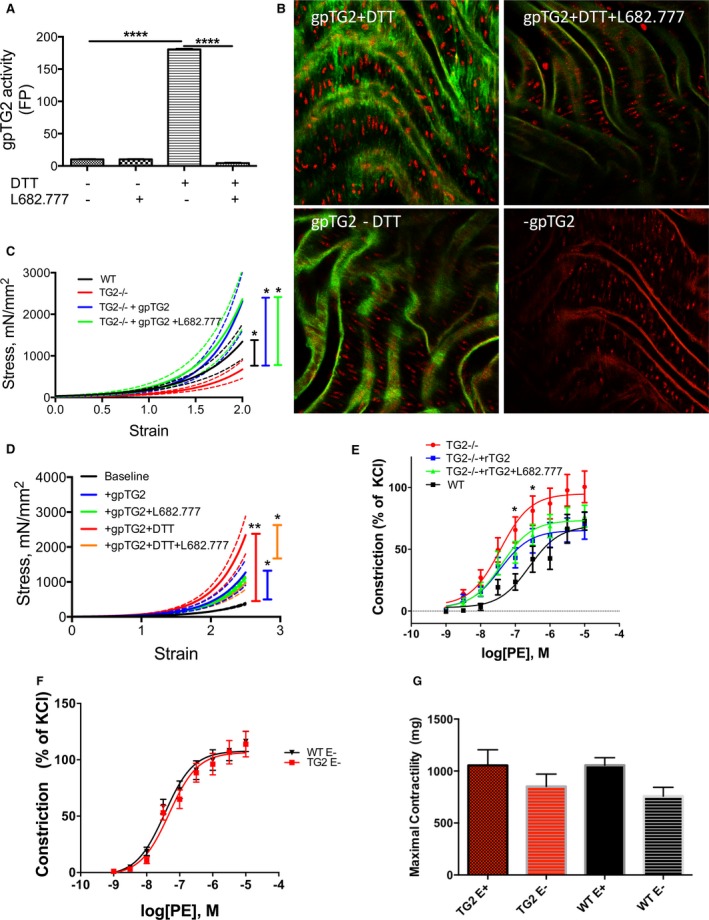
Methods and results: SMCs were isolated from the aortae of TG2-/- and wild-type (WT) mice. Cell adhesion was examined by using electrical cell-substrate impedance sensing and PicoGreen assay. Cell motility was examined using a Boyden chamber assay. Cell proliferation was examined by electrical cell-substrate impedance sensing and EdU incorporation assays. Cell micromechanics were studied using magnetic torsion cytometry and spontaneous nanobead tracer motions. Aortic mechanics were examined by tensile testing. Vasoreactivity was studied by wire myography. SMCs from TG2-/- mice had delayed adhesion, reduced motility, and accelerated de-adhesion and proliferation rates compared with those from WT. TG2-/- SMCs were stiffer and displayed fewer cytoskeletal remodeling events than WT. Collagen assembly was delayed in TG2-/- SMCs and recovered with adenoviral transduction of TG2. Aortic rings from TG2-/- mice were less stiff than those from WT; stiffness was partly recovered by incubation with guinea pig liver TG2 independent of crosslinking function. TG2-/- rings showed augmented response to phenylephrine-mediated vasoconstriction when compared with WT. In human coronary arteries, vascular media and plaque, high abundance of fibronectin expression, and colocalization with TG2 were observed.

Conclusions: TG2 modulates vascular function/tone by altering SMC contractility independent of its crosslinking function and contributes to vascular stiffness by regulating SMC proliferation and matrix remodeling.

Keywords: crosslinking; matrix remodeling; tissue transglutaminase; vascular biology; vascular disease; vascular smooth muscle; vascular stiffness; vessel.


Figures

Figure 1
Figure 1
TG2 regulates smooth muscle cell (SMC) function. SMCs were isolated from wild‐type (WT) and TG2−/− mouse aortae. A, Representative Western blot from 8 independent experiments showing that TG2 expression was present in WT SMCs but absent from TG2−/− SMCs; α‐actin was used as a loading control. B, Adhesion of TG2−/− SMCs on plastic, fibronectin, and collagen I supports was lower than that of WT SMCs by PicoGreen assay (*P<0.05, **P<0.01; 2‐way ANOVA with Bonferroni post hoc correction; n=7). C, Adhesion of TG2−/− SMCs was delayed on uncoated supports as examined by electrical cell‐substrate impedance sensing (ECIS). Data are shown as mean (solid line)±SE of measurement (broken lines); ***P<0.001 by Kruskal–Wallis test; n=6 for TG2−/− and WT SMCs, n=4 for cell‐free. D, EdU incorporation assay showed that proliferation of TG2−/− cells was elevated (*P<0.05; by Wilcoxon rank‐sum test; n=6 for WT, n=9 for TG2−/−). E, TG2−/− cells reached confluence earlier than WT cells and had a lower steady‐state resistance as examined by ECIS (**P<0.01; Kruskal–Wallis test; n=6 for TG2−/− and WT SMCs, n=4 for cell‐free condition). TG2 indicates tissue transglutaminase.
Figure 2
Figure 2
TG2 contributes to smooth muscle cell (SMC) micromechanics. A, TG2−/− SMCs showed strikingly lower cytoskeletal remodeling dynamics when compared with wild‐type (WT) SMCs (n=540 WT cells, 525 TG2−/− cells; 4 independent measurements; *P<0.05). B and C, Mean square displacements were significantly lower in TG2−/− cells at 10 s (B) and 300 s (C). D, TG2−/− SMCs showed diminished motility in response to 10% serum as chemoattractant in a Boyden chamber assay when compared with that of WT SMCs (*P<0.05; by Kruskal–Wallis test; n=9). E, Both storage (g′) and loss (g″) moduli of TG2−/− SMCs were markedly higher than those of WT SMCs over 5 decades of frequency (n=186 WT cells, 227 TG2−/− cells; 4 independent measurements). F, Overall cell stiffness was greater in TG2−/− cells than in WT cells. G, De‐adhesion from uncoated, fibronectin‐coated, and collagen‐coated cell culture dishes was faster in TG2−/− SMCs than in WT SMCs in response to treatment with 0.05% trypsin (**P<0.01 vs WT; # P<0.05 vs plastic WT by 2‐way ANOVA with Bonferroni post‐hoc correction; n=6). RFU indicates relative fluorescence units; TG2, tissue transglutaminase.
Figure 3
Figure 3
TG2 participates in collagen assembly. A, Collagen assembly was delayed in TG2−/− smooth muscle cells (SMCs; panels III, IV) when compared with that of WT SMCs (I, II). Adenoviral delivery of TG2 to TG2−/− SMCs (panels V, VI) recapitulated the WT time course for collagen assembly. Cell‐free samples did not show any assembled collagen fibers (panels VII, VIII). Images are representative of 6 independent experiments. B, Representative Western blot showing TG2 and α‐actin expression at days 1 and 3. TG2 indicates tissue transglutaminase; WT, wild type.
Figure 4
Figure 4
TG2 contributes to aortic modulus. A, Decellularized and (B) intact aortic segments from WT mice were significantly stiffer than those from TG2−/− mice; solid lines represent arithmetic mean and dotted lines represent SE of measurement (*P<0.05). C, Maximum strain at sample rupture was lower in the decellularized segments than in intact segments and similar between genotypes (*P<0.05). D, Maximum stress was higher in the decellularized segments than in intact aorta for the TG2−/− genotype but not the WT genotype. E, Lumen diameters were larger in decellularized segments than in intact segments for WT aorta (*P<0.05). F, Walls were significantly thinner in decellularized samples than in intact samples, in WT aorta (n=12 per group; *P<0.05). G, Pulse wave velocity (PWV)–mean arterial pressure (MAP) correlation was similar in 12‐ to 14‐week‐old WT and TG2−/− mice (n=8). H, Representative Western blot showing that TG2 protein was expressed in aorta from WT but not TG2−/− mice; GAPDH was used as loading control. TG2 indicates tissue transglutaminase; WT, wild type.
Figure 5
Figure 5
Exogenous TG2 affects vascular stiffness and vasoreactivity independent of crosslinking function. A, Activity of exogenous guinea pig TG2 (gpTG2) was examined by quantifying the crosslinking of FITC‐cadaverine and N,N′ dimethyl casein by fluorescence polarization. Dithiothreitol (DTT) activated and L682.777 inhibited gpTG2 (*P<0.05 by Kruskal–Wallis test; n=12 per group). (B) GpTG2 activity was examined by confocal microscopy in TG2−/− mouse aorta. GpTG2‐dependent incorporation of FITC‐cadaverine occurred in the presence of DTT and was inhibited by L682.777. Negligible FITC‐cadaverine incorporation was observed in samples lacking DTT and those lacking gpTG2 treatment. Images are representative of 8 independent experiments. C, Aortic modulus was examined by tensile testing; solid lines represent arithmetic mean and dotted lines represent SE of measurement. Incubation of TG2−/− mouse aorta with gpTG2 increased aortic stiffness to levels similar that of WT aorta both in the absence (blue) and presence (green) of TG2 inhibitor L682.777 (*P<0.05; n=8 per group). D, Incubation of rat aorta with DTT‐activated gpTG2 significantly increased stiffness (red). GpTG2 in the absence of DTT (blue) also resulted in a significant increase in stiffness when compared with baseline (black), but the magnitude of increase was reduced. L682.777 partially reversed the increase in stiffness in DTT‐activated gpTG2 (orange) and had no effect in the absence of DTT (green) n=8 per group; *P<0.05, **P<0.01. E, Vasoconstriction of aortic rings in response to increasing doses of phenylephrine (PE) was examined by wire myography. The response of rings from TG2−/− mice (red) was significantly left‐shifted compared with those from WT mice (black). Incubation of TG2−/− aortic rings with gpTG2 restored contractility toward that of WT in the absence (blue) and presence (green) of L682.777. n=8 per group; *P<0.05. (F) Endothelium‐denuded rings of TG2−/− and WT mice show similar responses to PE, suggesting an endothelial component to vascular responses. (G) Maximal contractility with KCl (60 mmol/L) is similar in intact TG2−/− and WT vessels and in denuded TG2−/− and WT vessels; denuded vessels had lower contractility than their endothelium‐intact counterparts. FITC indicates fluorescein isothiocyanate; TG2, tissue transglutaminase; WT, wild type.
Figure 6
Figure 6
TG2 and fibronectin colocalize in plaque in human coronary arteries. A 99‐feature human coronary artery tissue microarray (TMA) was used to examine the role of TG2 in human disease. A, A photograph of the TMA with hematoxylin & eosin (H&E) staining. B, Sample H&E and (C) smooth muscle actin stains of a single feature. D, TG2 expression was high in the vascular media but varied in plaque from very rare to highly abundant. E, Fibronectin expression was higher in plaque than in media. F, Colocalization of TG2 and fibronectin was more abundant in plaque than in media. G, TG crosslinking was similar in all samples. Representative immunofluorescence images in plaque (H) and vascular media (I; green=TG2, red=fibronectin, blue=nuclei); arrows indicate sample areas of colocalization. *P<0.05. TG2 indicates tissue transglutaminase.

References

    1. Zieman SJ, Melenovsky V, Kass DA. Mechanisms, pathophysiology, and therapy of arterial stiffness. Arterioscler Thromb Vasc Biol. 2005;25:932–943. - PubMed
    1. Vlachopoulos C, Aznaouridis K, Stefanadis C. Prediction of cardiovascular events and all‐cause mortality with arterial stiffness: a systematic review and meta‐analysis. J Am Coll Cardiol. 2010;55:1318–1327. - PubMed
    1. Lacolley P, Challande P, Osborne‐Pellegrin M, Regnault V. Genetics and pathophysiology of arterial stiffness. Cardiovasc Res. 2009;81:637–648. - PubMed
    1. Qiu H, Zhu Y, Sun Z, Trzeciakowski JP, Gansner M, Depre C, Resuello RR, Natividad FF, Hunter WC, Genin GM, Elson EL, Vatner DE, Meininger GA, Vatner SF. Short communication: vascular smooth muscle cell stiffness as a mechanism for increased aortic stiffness with aging. Circ Res. 2010;107:615–619. - PMC - PubMed
    1. Sehgel NL, Sun Z, Hong Z, Hunter WC, Hill MA, Vatner DE, Vatner SF, Meininger GA. Augmented vascular smooth muscle cell stiffness and adhesion when hypertension is superimposed on aging. Hypertension. 2015;65:370–377. - PMC - PubMed

MeSH terms

LinkOut - more resources