. 2017 Mar 7;114(10):E2046-E2052.
doi: 10.1073/pnas.1621248114. Epub 2017 Feb 3.

Multiple functional self-association interfaces in plant TIR domains

Affiliations

Multiple functional self-association interfaces in plant TIR domains

Xiaoxiao Zhang et al. Proc Natl Acad Sci U S A. .

Abstract

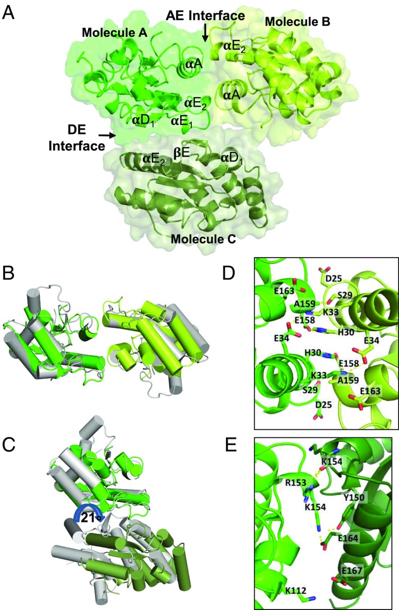
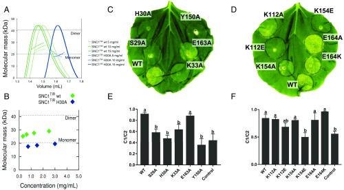
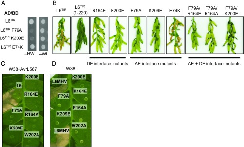
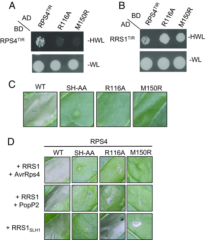
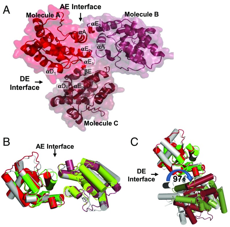
The self-association of Toll/interleukin-1 receptor/resistance protein (TIR) domains has been implicated in signaling in plant and animal immunity receptors. Structure-based studies identified different TIR-domain dimerization interfaces required for signaling of the plant nucleotide-binding oligomerization domain-like receptors (NLRs) L6 from flax and disease resistance protein RPS4 from Arabidopsis Here we show that the crystal structure of the TIR domain from the Arabidopsis NLR suppressor of npr1-1, constitutive 1 (SNC1) contains both an L6-like interface involving helices αD and αE (DE interface) and an RPS4-like interface involving helices αA and αE (AE interface). Mutations in either the AE- or DE-interface region disrupt cell-death signaling activity of SNC1, L6, and RPS4 TIR domains and full-length L6 and RPS4. Self-association of L6 and RPS4 TIR domains is affected by mutations in either region, whereas only AE-interface mutations affect SNC1 TIR-domain self-association. We further show two similar interfaces in the crystal structure of the TIR domain from the Arabidopsis NLR recognition of Peronospora parasitica 1 (RPP1). These data demonstrate that both the AE and DE self-association interfaces are simultaneously required for self-association and cell-death signaling in diverse plant NLRs.

Keywords: NLR; TIR domain; plant disease resistance; plant immunity; signaling by cooperative assembly formation.


Conflict of interest statement

The authors declare no conflict of interest.

Figures

Fig. 1.
Fig. 1.
The crystal structure of SNC1TIR reveals two self-association interfaces. (A) SNC1TIR crystal structure contains two major interfaces, involving predominantly αA and αE (AE) and αD and αE (DE) regions of the protein. The green and lime-colored SNC1TIR molecules are observed in the asymmetric unit and interact through the AE interface; the green molecule also interacts with a crystallographic symmetry-related molecule (forest colored) through the DE interface. (B) Superposition of the SNC1TIR (green and lime) and RPS4TIR (gray) AE-interface dimers; one chain in the pair was used for superposition. (C) As in B, but showing the superposition of SNC1TIR and L6TIR (gray) DE-interface dimers; note the ∼21° rotation at the DE interface between the two structures. (D) Residues that contribute to the buried surface in the AE-interface interactions in SNC1TIR are highlighted in stick representation. (E) As in D, showing the DE interface.
Fig. 2.
Fig. 2.
SNC1TIR self-association and signaling. (A) Solution properties of SNC1TIR (WT, wild-type) and SNC1TIR H30A analyzed by SEC-MALS. Green or blue peaks indicate the traces from the refractive index (RI) detector during SEC of SNC1TIR or its H30A mutant, respectively. The lines under the peaks correspond to the average molecular mass distributions across the peak (using equivalent coloring). (B) Molecular masses calculated from SAXS data for SNC1TIR (WT, wild-type; green diamonds) and SNC1TIR H30A (blue diamonds), calculated from static samples at discrete concentrations between 3 and 0.25 mg/mL. Dotted lines indicate the theoretical monomeric and dimeric masses. (C–F) In planta mutational analysis of SNC1TIR. (C and D) Autoactive phenotype of SNC1TIR (residues 1–226; WT, wild-type) and the corresponding mutants upon Agrobacterium-mediated transient expression in N. benthamiana leaves. Each construct was coexpressed with the virus-encoded suppressor of gene silencing P19 (33). Photos were taken 5 d after infiltration. (E and F) Ion-leakage measurement of the infiltrated leaves as shown in C and D. Each construct was expressed in independent leaves. Leaf disk samples were collected 2 d after infiltration and incubated in Milli-Q water. C1 corresponds to the ions released in solution 24 h after sampling. C2 corresponds to the total ion contents in the sample (see SI Appendix, Methods for details). Ion leakage was calculated as C1/C2 ratio. N. benthamiana leaves expressing P19 only were used as control. Error bars show SE of means. Statistical differences, calculated by one-way ANOVA and multiple comparison with the control, are indicated by letters.
Fig. 3.
Fig. 3.
Mutations in both AE and DE interfaces affect L6TIR self-association and autoactivity and full-length L6 effector-dependent and effector-independent cell-death signaling. (A) Mutations in the AE interface disrupt L6TIR self-association in yeast. Growth of yeast cells expressing GAL4-BD and GAL4-AD fusions of L6TIR (residues 29–233) or L6TIR mutants on nonselective media lacking tryptophan and leucine (−WL) or selective media additionally lacking histidine (−HWL). (B) Mutations in the AE interface disrupt L6TIR signaling activity in planta. Cell-death signaling activity of L6TIR (residues 1–233) mutants fused to yellow fluorescent protein (YFP), 12 d after agroinfiltration in flax plants. The truncated L6 TIR domain (residues 1–220) was used as a negative control (9). Agrobacterium cultures carrying L6TIR mutants were adjusted to OD1. (CD) Representative cell-death activity of L6 (C) and L6MHV (D) mutants, fused to YFP, 3 d after agroinfiltration in wild-type tobacco W38 or transgenic tobacco W38 carrying AvrL567, respectively. Agrobacterium cultures carrying L6 and L6MHV mutant were adjusted to OD 0.5.
Fig. 4.
Fig. 4.
Mutations in both AE and DE interfaces affect RPS4TIR self-association and autoactivity and full-length RPS4 effector-dependent and effector-independent cell-death signaling. (A) Mutations in the DE interface disrupt RPS4TIR self-association in yeast. Growth of yeast cells expressing GAL4-BD fusion and GAL4-AD fusion of RPS4TIR (residues 1–183) or RPS4TIR mutants on nonselective media lacking tryptophan and leucine (−WL) or selective media additionally lacking histidine (−HWL). (B) Mutations in the DE interface do not affect RPS4TIR interaction with RRS1TIR. Growth of yeast cells coexpressing GAL4-BD fusion of RPS4TIR or RPS4TIR mutants and GAL4-AD fusion of RRS1TIR (residues 1–185) on −WL or −HWL media. (C) Mutations in the DE interface disrupt RPS4TIR signaling activity in planta. Cell-death signaling activity of RPS4TIR (WT, wild-type) and its mutants fused to C-terminal 6xHA tags, 3 d after agroinfiltration in tobacco. (D) Representative cell-death activity of full-length RPS4 (WT, wild-type) and its mutants fused to C-terminal 3xHA tags, upon agro-mediated transient coexpression with RRS1 and corresponding effectors (AvrRps4 or PopP2), or with RRS1SLH1 mutant in W38 tobacco. Agrobacterium cultures were adjusted to OD 0.1. Photos were taken 5 d after agroinfiltration.
Fig. 5.
Fig. 5.
The AE and DE interface in the crystal structure of RPP1TIR. (A) Ribbon representation of the RPP1 crystal structure and the AE and DE interfaces, with molecules sharing the AE interface colored red and raspberry and the DE interface, red and ruby. (B) Comparison of the AE interface from the RPS4TIR (gray), SNC1TIR (green and lime), and RPP1TIR (red and raspberry) with the chains on the Left superimposed, highlighting the strong structural conservation of the interface. (C) Comparison of the DE interface from the L6TIR (gray), SNC1TIR (green and forest), and RPP1TIR (red and ruby) structures; only the chains at the Top are superimposed, highlighting the rotation observed at the DE interface in these crystal structures.

Comment in

  • Two-faced TIRs trip the immune switch.
    Jones JD, Banfield MJ. Jones JD, et al. Proc Natl Acad Sci U S A. 2017 Mar 7;114(10):2445-2446. doi: 10.1073/pnas.1700954114. Epub 2017 Feb 27. Proc Natl Acad Sci U S A. 2017. PMID: 28242711 Free PMC article. No abstract available.

References

    1. Dodds PN, Rathjen JP. Plant immunity: Towards an integrated view of plant-pathogen interactions. Nat Rev Genet. 2010;11(8):539–548. - PubMed
    1. Bentham A, Burdett H, Anderson PA, Williams SJ, Kobe B. Animal NLRs provide structural insights into plant NLR function. Ann Bot. August 24, 2016 doi: 10.1093/aob/mcw171. - DOI - PMC - PubMed
    1. Duxbury Z, et al. Pathogen perception by NLRs in plants and animals: Parallel worlds. BioEssays. 2016;38(8):769–781. - PubMed
    1. van der Biezen EA, Jones JD. The NB-ARC domain: A novel signalling motif shared by plant resistance gene products and regulators of cell death in animals. Curr Biol. 1998;8(7):R226–R227. - PubMed
    1. McHale L, Tan X, Koehl P, Michelmore RW. Plant NBS-LRR proteins: Adaptable guards. Genome Biol. 2006;7(4):212. - PMC - PubMed

Publication types

MeSH terms