. 2017 Apr 20;45(7):3967-3984.
doi: 10.1093/nar/gkx073.

Arm-specific cleavage and mutation during reverse transcription of 2΄,5΄-branched RNA by Moloney murine leukemia virus reverse transcriptase

Affiliations

Arm-specific cleavage and mutation during reverse transcription of 2΄,5΄-branched RNA by Moloney murine leukemia virus reverse transcriptase

Jessica Döring et al. Nucleic Acids Res. .

Abstract

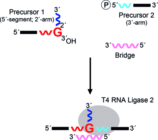
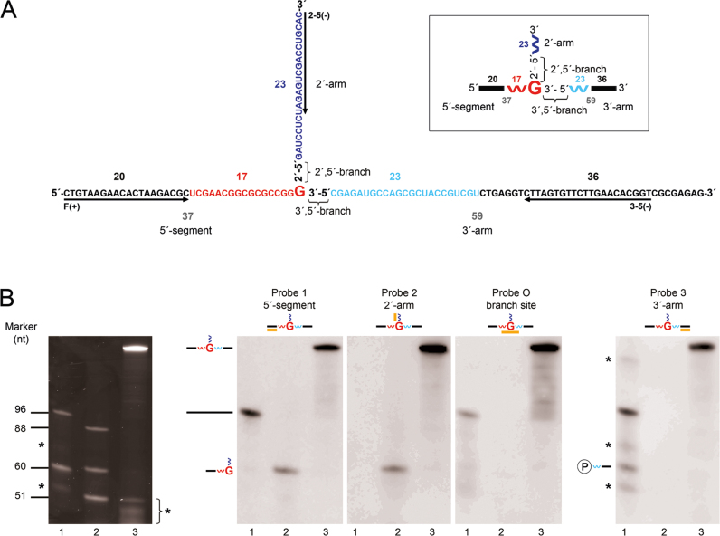
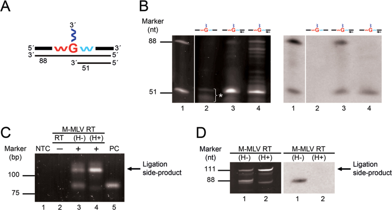
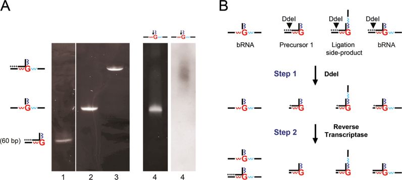
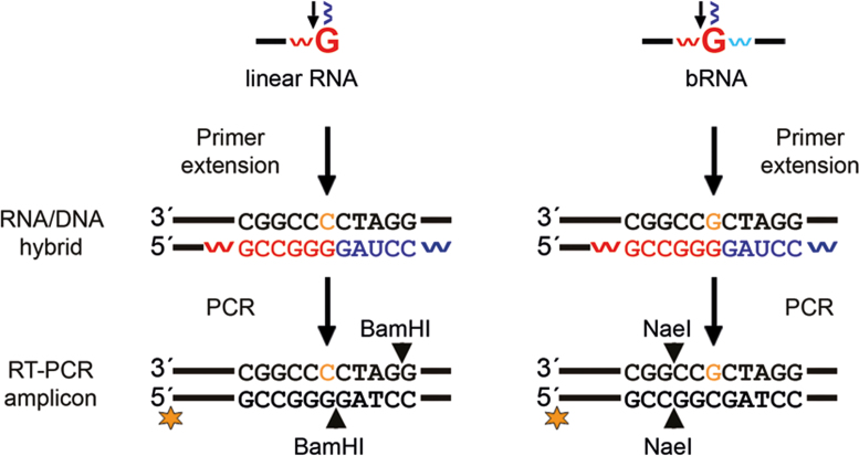
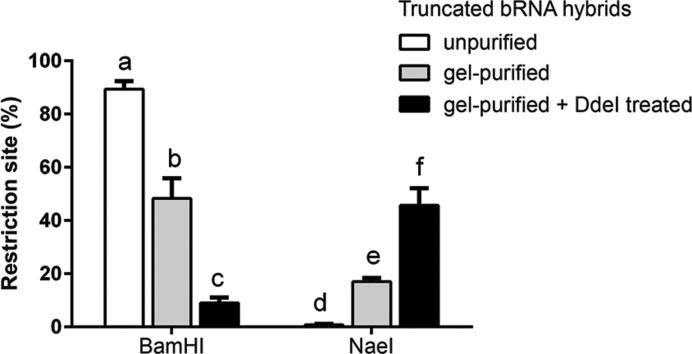
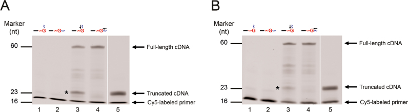
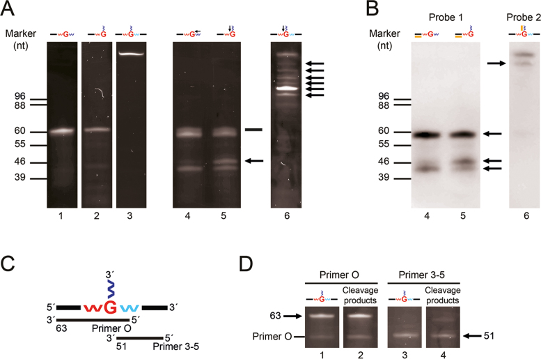
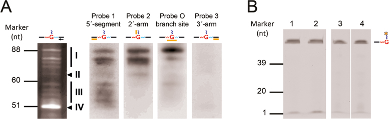
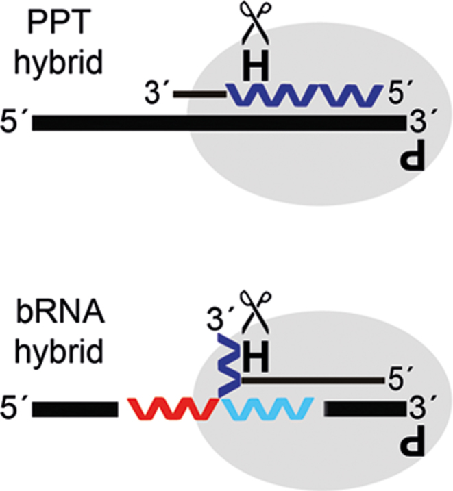
Branchpoint nucleotides of intron lariats induce pausing of DNA synthesis by reverse transcriptases (RTs), but it is not known yet how they direct RT RNase H activity on branched RNA (bRNA). Here, we report the effects of the two arms of bRNA on branchpoint-directed RNA cleavage and mutation produced by Moloney murine leukemia virus (M-MLV) RT during DNA polymerization. We constructed a long-chained bRNA template by splinted-ligation. The bRNA oligonucleotide is chimeric and contains DNA to identify RNA cleavage products by probe hybridization. Unique sequences surrounding the branchpoint facilitate monitoring of bRNA purification by terminal-restriction fragment length polymorphism analysis. We evaluate the M-MLV RT-generated cleavage and mutational patterns. We find that cleavage of bRNA and misprocessing of the branched nucleotide proceed arm-specifically. Bypass of the branchpoint from the 2΄-arm causes single-mismatch errors, whereas bypass from the 3΄-arm leads to deletion mutations. The non-template arm is cleaved when reverse transcription is primed from the 3΄-arm but not from the 2΄-arm. This suggests that RTs flip ∼180° at branchpoints and RNases H cleave the non-template arm depending on its accessibility. Our observed interplay between M-MLV RT and bRNA would be compatible with a bRNA-mediated control of retroviral and related retrotransposon replication.


Figures

Figure 1.
Figure 1.
Scheme of the splinted-ligation method in bRNA construction. In this method, a 2΄-5΄ linked ribo-guanosine (G)-nucleoside in an RNA strand containing the 5΄-segment and 2΄-arm (precursor 1) is transformed into a branchpoint nucleotide by ligation to an RNA strand representing the 3΄-arm (precursor 2). To do so, the two precursors are hybridized partially to a complementary RNA bridge. In this way, the 5΄-phosphate of precursor 2 is brought close to the free 3΄-hydroxyl of the 2΄-5΄ linked nucleoside of precursor 1. The two oligonucleotides are then joined by T4 RNA Ligase 2. Red, blue, and pink symbols ‘w’ represent RNA; the black line represents DNA. The 2΄-5΄ linked ribo-G-nucleoside in precursor 1 at nucleotide (nt) position 37 is highlighted. Nucleic acids downstream of a 2΄-5΄ linkage are plotted vertically in linear and branched oligonucleotides.
Figure 2.
Figure 2.
Branched RNA oligonucleotide and site-specific detection by probe hybridization. (A) Sequence and length of the bRNA construct. The bRNA is composed of a 37-mer 5΄-segment, a 23-mer 2΄-arm, and a 59-mer 3΄-arm. The branchpoint nucleotide guanosine (branch guanosine) at nt position 37 is highlighted. The 2΄,5΄-phosphodiester bond of the branch guanosine is designated as 2΄,5΄-branch and its 3΄,5΄-phosphodiester bond as 3΄,5΄-branch. Red and blue nucleotides represent RNA; black nucleotides represent DNA. Numbers refer to the length in nucleotides of the respective nucleic acid. The orientation of the shown bRNA construct was defined as ‘sense’. Primer binding sites are indicated by arrows. The primer with the suffix (+) is the forward primer and those with the (−) are the reverse primers. The primers 2–5 and 3–5 were used to prime DNA synthesis from the 2΄- and 3΄-arm, respectively. A schematic presentation of the constructed bRNA oligonucleotide is boxed in black. (B) Site-specific probes used in hybridization analyses. Lane 1: 96 nucleotides (nt) long oligonucleotide with the sequence of the bRNA construct without 2΄-arm and precursor 2 (59 nt). Lane 2: oligonucleotide representing full-length cDNA through the 3΄,5-branch (88 nt), precursor 1 (60 nt), and oligonucleotide representing cDNA until the 3΄,5΄-branch (51 nt). Lane 3: negative control for primer extension reaction using bRNA and reverse primer 3–5. Due to its unusual shape, the bRNA oligonucleotide exhibits an anomalous electrophoretic mobility in polyacrylamide gels and migrates more slowly than the corresponding linear oligonucleotide (oligonucleotide containing the 5΄-segment and 3΄-arm, 96 nt) (85). Samples were loaded on a 15% denaturing polyacrylamide gel (left), blotted and corresponding blots (right) were hybridized with site-specific probes (probes 1–3, probe O) as indicated. Probe 1 is specific for the 5΄-segment of bRNA, probe 2 for the 2΄-arm of bRNA, probe O for the overlapping branch site of bRNA, and probe 3 for the 3΄-arm of bRNA. Probe-target regions are plotted in pictograms for each blot, with probes shown in orange. Hybridizing oligonucleotides from lanes 1–3 are schematically presented on the left of the blots. By-products in solid-phase synthesis of oligonucleotides loaded in lane 1 and of reverse primer 3–5 loaded in lane 3 are labeled with asterisks.
Figure 3.
Figure 3.
Detection of full-length cDNA through the 3΄,5΄-branch and truncated cDNA until the 3΄,5΄-branch. (A) Schematic presentation of full-length and truncated cDNA generated by primer extension from the 3΄-arm of our bRNA construct. Numbers below lines refer to the lengths in nucleotides of the two cDNAs. (B) Analysis of primer extension products synthesized by M-MLV RT (H−) and (H+) from the 3΄-arm by probe hybridization. Lane 1: oligonucleotide representing full-length cDNA through the 3΄,5΄-branch (88 nt) and oligonucleotide representing truncated cDNA until the 3΄,5΄-branch (51 nt). Lane 2: negative control for primer extension reaction. Asterisk indicates by-products in solid-phase synthesis of reverse primer 3–5. Lanes 3 and 4: primer extension reactions from the 3΄-arm of bRNA using M-MLV RT (H−) and (H+), respectively. The black arrow in the pictogram shows the primer-target region and direction of primer extension. Oligonucleotides from lane 1 were used as size markers for gel electrophoresis and as positive hybridization controls. Samples were separated on a 15% denaturing polyacrylamide gel (left), blotted and hybridized with probe tc3-5 (right). This probe is specific for full-length cDNA through the 3΄,5΄-branch and truncated cDNA until the 3΄,5΄-branch. (C) Detection of full-length cDNA through the 3΄,5΄-branch of our bRNA construct by RT-PCR analysis. One fmol of oligonucleotide corresponding to full-length cDNA through the 3΄,5΄-branch was used as a positive control for PCR. Lane 1: no template control (NTC) for PCR. Lane 2: negative control for RT-PCR (no RT was added). Lanes 3 and 4: RT-PCR amplicons. Lane 5: positive control (PC). The RT-PCR product corresponding to the unwanted ligation side-product is noted. Samples were loaded on a 4% agarose gel. (D) Analysis of RT-PCR amplicons by probe hybridization. Sixty ng of column-purified RT-PCR amplicons (lanes 1 and 2) shown in panel (C) were separated on a 15% denaturing polyacrylamide gel (left), blotted and hybridized with probe flc3-5 (right). This probe is specific for full-length cDNA through the 3΄,5΄-branch. The ligation side-product is noted as in panel (C). Colors as in Figure 1.
Figure 4.
Figure 4.
Depletion of full-length hybrids from bRNA. (A) Gel purification of truncated bRNA hybrids. Lane 1: hybrid between precursor 1 and full-length cDNA. Lane 2: hybrid between bRNA and truncated cDNA until the 2΄,5΄-branch. Lane 3: hybrid between bRNA and full-length cDNA. Lane 4: products of primer extension reaction from the 2΄-arm of bRNA using M-MLV RT (H−). Hybrids from lanes 1–3 are schematically presented on the left of the gel. Pluses in pictograms indicate the forward primer binding site (PBS) of full-length cDNA. Hybrids (lanes 1–3) were used as size markers for electrophoresis on a 12% native polyacrylamide gel. The primer extension reaction from lane 4 was blotted and hybridized with probe flc2-5 (right). This probe is specific for full-length cDNA through the 2΄-5΄-linkage of linear RNA and bRNA. Arrows in pictograms as in Figure 3B. (B) Scheme showing truncation of the DNA duplex of full-length hybrids by restriction digestion. Truncated and full-length bRNA hybrids, as well as full-length linear RNA hybrids are presented schematically at the top. Pluses in pictograms as in panel (A). The DdeI restriction site in the DNA duplex of full-length hybrids is shown. DdeI restriction digestion removes the forward PBS of full-length cDNAs and truncates templates (step 1). In the next round of primer extension from the 2΄-arm, M-MLV RT (H-) generates truncated and full-length cDNA from bRNA, and truncated cDNAs from truncated templates (step 2). Colors as in Figure 1.
Figure 5.
Figure 5.
RT-PCR amplicons from linear RNA and bRNA can be distinguished by restriction digestion. Linear and bRNA templates are presented schematically at the top. Arrows in pictograms as in Figure 3B. Orange nucleotides indicate the incorporated nucleotide by M-MLV RT (H−) opposite to the 2΄-5΄ linked ribo-G-nucleoside in linear RNA and in bRNA. They can be identified in RT-PCR amplicons using terminal-restriction fragment length polymorphism (T-RFLP) analysis. For this purpose, the forward primer was labeled at the 5΄-end with the fluorophore cy3 (indicated by a star). Colors as in Figure 1.
Figure 6.
Figure 6.
Frequency distribution of BamHI and NaeI restriction sites in RT-PCR amplicons. T-RFLP patterns of BamHI and NaeI digested RT-PCR amplicons obtained from unpurified, gel-purified or gel-purified and DdeI treated truncated bRNA hybrids shown in Supplementary Figure S2 were evaluated quantitatively. Error bars above columns indicate standard deviations from three independent experiments. Columns headed by different letters are significantly different from each other at P < 0.05 according to analysis of variance (ANOVA) followed by Holm-Sidak's multiple comparison test.
Figure 7.
Figure 7.
Effect of the 2΄-5΄ linked ribo-G-nucleoside in precursor 1 on DNA synthesis by M-MLV RT (H+) and (H−). (A) DNA synthesis catalyzed by the wild-type RT. Primer extension reactions were primed with the 5΄-cy5-labeled reverse primer 2_5. Lanes 1 and 2: negative controls for primer extension reaction on precursor 1 and control template with only 3΄-5΄ linkages, respectively. Lanes 3 and 4: primer extension reactions on precursor 1 and control template, respectively. Lane 5: primer extension reaction on oligonucleotide representing the 2΄-arm using M-MLV RT (H−). Colors as in Figure 1 and arrows in pictograms as in Figure 3B. Samples were separated on a 15% denaturing polyacrylamide gel. Full-length cDNA and truncated cDNA until the 2΄-5΄ linkage, as well as the cy5-labeled primer are noted. (B) DNA synthesis catalyzed by the RNase H-deficient RT. Legend as in panel (A). The cDNA that stops at the 2΄,5΄-linkage is labeled with an asterisk.
Figure 8.
Figure 8.
Characterizing RNA cleavage products generated by M-MLV RT RNase H during DNA synthesis from the 2΄-arm. (A) Typical RNA cleavage patterns of oligonucleotides containing either a 3΄-5΄ linkage (control template), a 2΄-5΄ linkage (precursor 1), or a branched nucleotide (bRNA) at nt position 37. Lanes 1–3: negative controls for RNase H cleavage of the control template, precursor 1, and bRNA, respectively. Lanes 4–6: RNase H cleavages of the control template, precursor 1 and bRNA, respectively, during DNA synthesis from the 2΄-arm. Arrows in pictograms as in Figure 3B. Samples were electrophoresed on a 15% denaturing polyacrylamide gel. Pause-related cleavage products are labeled with an arrow, and full-length cDNA from linear templates is indicated by a black bar (B) Analysis of RNA cleavage products by probe hybridization. RNA cleavage patterns (lanes 4–6) shown in panel (A), were blotted and hybridized with either probe 1 (lanes 4 and 5) or probe 2 (lane 6) as indicated. Probe specificities and probe-target regions as in Figure 2B. Hybridizing cleavage products are labeled with an arrow. (C) Scheme showing the primer extension method. Branched RNA and cDNAs produced with the reverse primers O and 3–5 are presented. Numbers below lines refer to the lengths in nucleotides of the generated cDNAs. (D) Primer extension analysis of bRNA and RNA cleavage products using M-MLV RT (H−). Lanes 1 and 2: primer extension reactions with primer O on bRNA and RNA cleavage products, respectively. The primer band of primer O is noted. Lanes 3 and 4: primer extension reactions with primer 3–5 on bRNA and RNA cleavage products, respectively. Samples were analyzed on a 15% denaturing polyacrylamide gel. Complementary DNAs generated from bRNA and RNA cleavage products are labeled with arrows, where the numbers indicate their lengths in nucleotides. Colors as in Figure 1.
Figure 9.
Figure 9.
Characterizing RNA cleavage products generated by M-MLV RT RNase H during DNA synthesis from the 3΄-arm. (A) Hybridization analyses of cleavage products. Typical RNA cleavage pattern of the bRNA construct separated on a 15% denaturing polyacrylamide gel is shown on the left (same gel as in Figure 3B, lane 4). RNA cleavage patterns were blotted and corresponding blots (right) were hybridized with specific probes (probes 1–3, probe O) as indicated. Probe specificities and probe-target regions as in Figure 2B. Products were grouped into four clusters (I–IV) according to their hybridization with probes as indicated on the right of the gel. Cleavage products of cluster I hybridized with probes 1, 2 and O, cleavage product of cluster II with probes 1 and 2, cleavage products of cluster III with probe 1, and product of cluster IV was identified as truncated cDNA until the 3΄,5΄-branch (see Figure 3B, lane 4). (B) Primer extension analyses on the 3΄-fluorescein-labeled precursor 1 using M-MLV RT (H+). Lanes 1 and 3: negative controls for primer extension reaction, respectively. Lane 2: primer extension reaction using reverse primer 3–5. Lane 4: primer extension reaction using reverse primer 3–5 and bRNA. The 3΄-fluorescein-labeled precursor 1 is schematically presented on the right, where the star indicates the position of the fluorophore. Samples were separated on a 15% denaturing polyacrylamide gel. The bands migrating at the bottom of the gel represented the loading dye. Colors as in Figure 1.
Figure 10.
Figure 10.
Flipped bound state of RT on polypurine tract (PPT) and bRNA hybrids. The relative positions of the RNase H and DNA polymerase catalytic sites of RT bound to the PPT and bRNA hybrid are presented. H with the scissor and the rotated P on the enzyme represent the active RNase H and inactive DNA polymerase catalytic site, respectively. The non-template strand is coloured in dark blue, otherwise colors as in Figure 1.

Similar articles

Cited by

References

    1. Herschhorn A., Hizi A.. Retroviral reverse transcriptases. Cell. Mol. Life Sci. 2010; 67:2717–2747. - PMC - PubMed
    1. Le Grice S.F.J., Nowotny M.. Nucleic Acid Polymerases. 2014; 30:Berlin Heidelberg, NY: Springer-Verlag; 189–214.
    1. Krug M.S., Berger S.L.. Ribonuclease H activities associated with viral reverse transcriptases are endonucleases. Proc. Natl. Acad. Sci. U.S.A. 1989; 86:3539–3543. - PMC - PubMed
    1. DeStefano J.J., Buiser R.G., Mallaber L.M., Bambara R.A., Fay P.J.. Human immunodeficiency virus reverse transcriptase displays a partially processive 3΄ to 5΄ endonuclease activity. J. Biol. Chem. 1991; 266:24295–24301. - PubMed
    1. Schultz S.J., Champoux J.J.. RNase H activity: structure, specificity, and function in reverse transcription. Virus Res. 2008; 134:86–103. - PMC - PubMed

Publication types

MeSH terms

LinkOut - more resources