Review
. 2017 Feb;42(2):118-126.
doi: 10.1016/j.jhsa.2016.11.025.

Elbow Instability: Anatomy, Biomechanics, Diagnostic Maneuvers, and Testing

Affiliations
Review

Elbow Instability: Anatomy, Biomechanics, Diagnostic Maneuvers, and Testing

Lauren E Karbach et al. J Hand Surg Am. 2017 Feb.

Abstract

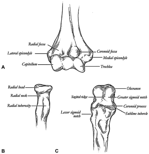
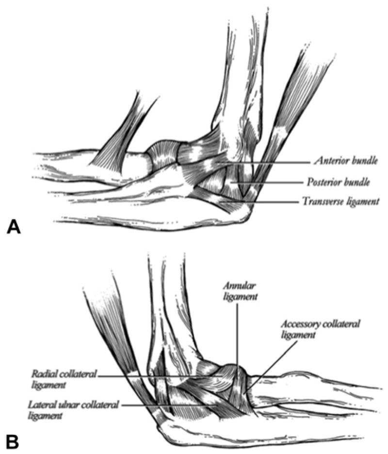
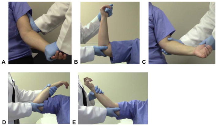
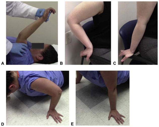
The elbow comprises a complex of bony and ligamentous stabilizers that provide both primary and secondary constraints to elbow instability. Through trauma and overuse, classic instability patterns arise by loss of these important stabilizers. The diagnosis of elbow instability can made using specific examination maneuvers and testing to diagnose the clinical pattern. This article reviews the elbow's unique anatomy and biomechanical characteristics and these are applied when reviewing the maneuvers and testing used to diagnose elbow instability.

Keywords: Valgus instability; elbow biomechanics; posterolateral rotatory instability; varus posteromedial rotatory instability.


Figures

FIGURE 1
FIGURE 1
Osseous anatomy of the elbow. A Distal humerus. B Proximal radius. C Proximal ulna. (From Total elbow arthroplasty design. In: Williams GR, Yamaguchi K, Ramsey ML, et al, eds. Shoulder and Elbow Arthroplasty. Philadelphia: Lippincott Williams & Wilkins; 2005:301. Reprinted with permission.)
FIGURE 2
FIGURE 2
Medial and lateral ligamentous stabilizers of the elbow. A MCL complex. B LCL complex. (From Total elbow arthroplasty design. In: Williams GR, Yamaguchi K, Ramsey ML, et al, eds. Shoulder and Elbow Arthroplasty. Philadelphia: Lippincott Williams & Wilkins; 2005:303. Reprinted with permission.)
FIGURE 3
FIGURE 3
Diagnostic maneuvers for valgus elbow instability. A Valgus stress test. The elbow is flexed to 20°, the forearm is supinated, and a valgus stress is applied across the elbow. A positive test elicits pain over the medial ligaments or widening of the medial joint line. B Milking maneuver. The elbow is flexed to greater than 90° and a valgus stress is applied through the elbow by pulling on the patient’s thumb. C Modified milking maneuver. The arm is adducted and externally rotated, the patient’s elbow is flexed to 70°, and a valgus stress is applied through the thumb. D, E Moving valgus stress test. The patient’s shoulder is abducted and externally rotated while the elbow is ranged from maximal flexion to 30° flexion as a valgus force is applied. A patient with valgus instability will experience medial elbow pain or apprehension in the 70° to 120° flexion range.
FIGURE 4
FIGURE 4
PRLI tests. A Lateral pivot-shift test. The arm is raised above the head of the supine patient, the forearm is supinated, and a valgus and axial force is applied as the elbow is flexed. A “dimple” proximal to the radial head will be seen at 40° of flexion that produces a clunk with increased flexion. B, C. Chair push-up test. In a seated patient, the elbows are flexed to 90° and the forearms supinated. The patient then pushes up on the arms of the chair and extends the elbows. D, E Prone push-up test. The patient assumes a push-up position and supinates the forearm, then pushes up from 90° elbow flexion. A positive test will cause radial head subluxation or pain in a patient with PLRI.
FIGURE 5
FIGURE 5
Table-top relocation test for PLRI. A First component. The patient’s hand is supinated and grasps the edge of a table, then applies an axial force and flexes the elbow. The patient will have pain or radial head subluxation at approximately 40° of flexion. B Second component. From the starting position, the examiner applies force upon the radial head as the patient flexes the elbow. This prevents radial head subluxation and prevents pain. C Third component. The examiner then removes his or her thumb, which causes recurrence of the patient’s pain.
FIGURE 6
FIGURE 6
Gravity-assisted varus stress test for VPMI. A, B The patient’s arm is abducted to 90° in neutral rotation as the elbow is flexed and extended, allowing gravity to apply a varus stress.

References

    1. Hausman MR, Lang P. Examination of the elbow: current concepts. J Hand Surg Am. 2014;39(12):2534–2541. - PubMed
    1. Bryce CD, Armstrong AD. Anatomy and biomechanics of the elbow. Orthop Clin North Am. 2008;39(2):141–154. v. - PubMed
    1. Morrey BF, An KN. Stability of the elbow: osseous constraints. J Shoulder Elbow Surg. 2005;14(1 Suppl S):174S–178S. - PubMed
    1. Ahmed I, Mistry J. The management of acute and chronic elbow instability. Orthop Clin North Am. 2015;46(2):271–280. - PubMed
    1. Safran MR, Baillargeon D. Soft-tissue stabilizers of the elbow. J Shoulder Elbow Surg. 2005;14(1 Suppl S):179S–185S. - PubMed

MeSH terms

LinkOut - more resources