Review
. 2017 Feb:16:41-50.
doi: 10.1016/j.ebiom.2017.01.042. Epub 2017 Jan 31.

Memory B Cells are Major Targets for Effective Immunotherapy in Relapsing Multiple Sclerosis

Affiliations
Review

Memory B Cells are Major Targets for Effective Immunotherapy in Relapsing Multiple Sclerosis

David Baker et al. EBioMedicine. 2017 Feb.

Abstract

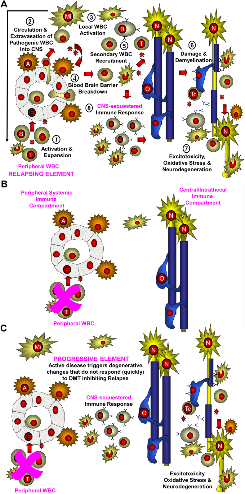
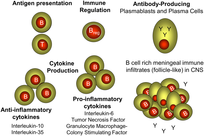
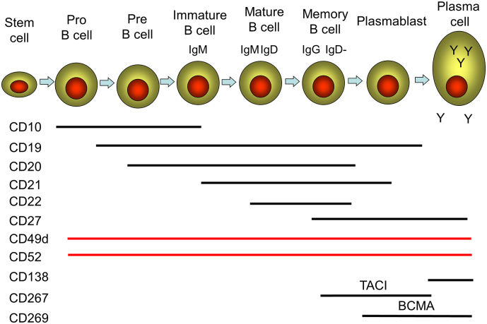
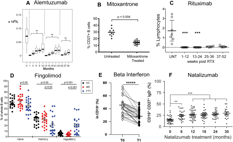
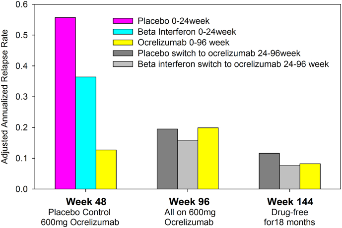
Although multiple sclerosis (MS) is considered to be a CD4, Th17-mediated autoimmune disease, supportive evidence is perhaps circumstantial, often based on animal studies, and is questioned by the perceived failure of CD4-depleting antibodies to control relapsing MS. Therefore, it was interestingly to find that current MS-treatments, believed to act via T cell inhibition, including: beta-interferons, glatiramer acetate, cytostatic agents, dimethyl fumarate, fingolimod, cladribine, daclizumab, rituximab/ocrelizumab physically, or functionally in the case of natalizumab, also depleted CD19+, CD27+ memory B cells. This depletion was substantial and long-term following CD52 and CD20-depletion, and both also induced long-term inhibition of MS with few treatment cycles, indicating induction-therapy activity. Importantly, memory B cells were augmented by B cell activating factor (atacicept) and tumor necrosis factor (infliximab) blockade that are known to worsen MS. This creates a unifying concept centered on memory B cells that is consistent with therapeutic, histopathological and etiological aspects of MS.

Keywords: Autoimmunity; Disease modifying treatment; Immunotherapy; Memory B cell; Multiple sclerosis.


Figures

Fig. 1
Fig. 1
Two immune-compartment model of multiple sclerosis. The initial trigger of the lesions is due to: (a) peripheral sensitization due to molecular mimicry or another event in the lymph node (outside-in) or (b) oligodendrocyte damage leading to liberation of antigen proteins or peptides that exit via the glymphatics to draining lymph nodes (inside-out) where autoreactive lymphocytes are sensitized. A. 1. Primed T and B cells are generated and travel round the body. 2. Immune cells enter into the CNS. 3. Following recognition of a target presented by a perivascular microglial (Mi) cell there is local activation of the infiltrating lymphocytes. 4. Cytokine release occurs to activate the blood brain barrier to express adhesion molecules. 5. A second wave of influx of T cells, B cells and monocytes enters the CNS. 6. These cause damage to the oligodendrocyte (O) via release of antibodies, and soluble products and possibly by direct killing by cytotoxic T cells (Tc). 7. Demyelinated nerves (N) have an elevated energy requirement to maintain neurotransmission. These are vulnerable to excitotoxic and other damage elements such as by activated microglial cells, and B cell products. 8. Microglial and B cells are sequestered into CNS compartment. B. Current DMD prevent entry of the peripheral adaptive immune cells into the CNS. This will block relapsing disease allowing natural repair mechanisms to act and induce a long-term status of no evidence of disease activity. C. These events produce an innate inflammatory environment formed from glial cells and adaptive immune niches, such as B cell infiltrates are created within the CNS. These may not responsive to peripheral immune control and may allow neurodegeneration and accumulation of disability to continue in the absence of active lesion formation.
Fig. 2
Fig. 2
Potential B cell functions in multiple sclerosis. B cells can exhibit a variety of different functions (bold) that may influence MS, both in the periphery and in the CNS where follicle-like structures accumulate during MS.
Fig. 3
Fig. 3
B cell lineage and surface marker expression. A diagram of a simplified development and differentiation pathway of the B cell lineage showing some distinguishing surface markers including ATCI (transmembrane activator and CAML interactor) protein and BCMA (B cell maturation antigen). The memory B cell subset is heterogeneous and includes both unswitched (CD19 +, CD27 +, IgM +, IgD +) and class switched (CD19 +, CD27 +, IgM −, IgD −) memory B cells, which were not individually reported in some of the studies analysed.
Fig. 4
Fig. 4
Active DMD in MS physically or functionally deplete memory B cell activity. Depletion of CD19 +, CD27 + memory B cells following treatment of MS with: (A) alemtuzumab (Thompson et al., 2010). The results show the depletion of the absolute numbers of memory B cells over three treatment cycles. **P < 0.01. Reproduced by permission from Springer Science: J. Clinical Immunology. DOI: 10.1007/s10875-009-9327-3. (B) Mitoxantrone (Duddy et al., 2007). The results show percentage reduction in the individual levels of CD19, CD27 memory B cell before (untreated) and one month after treatment. Reproduced by permission American Association of Immunology: Journal of Immunology. DOI: 10.4049/jimmunol.140011845. (C) Rituximab (Palanichamy et al., 2014). The results show the percentage of memory B cells (CD19, CD27, IgD −) in blood before, in untreated (UNT) individuals, and at different time points after rituximab (RTX). ***P < 0.0001. Reproduced by permission American Association of Immunology: Journal of Immunology. DOI: 10.4049/jimmunol.140011845. (D) Fingolimod (Grützke et al., 2015). The results are individual percentage of CD38, CD27 −, CD24 − mature/naïve cells (black); CD38 −, CD24 +, CD27 + memory B cells, CD38 +, CD27 −, CD24 +, CD5 + regulatory B cells in healthy controls (HC. Black dots) and people with multiple sclerosis who were untreated (MS. Red dots) or treated with oral fingolimod (FTY. Blue dots). *P < 0.05. Reproduced under creative commons license CC BY-NC-ND. DOI: 10.1002/acn3.155. (E) Beta interferon (Rizzo et al., 2016). The results show a reduction in the individual percentage of CD19 +, CD27 + B memory cells before (T0) and one month (T1) after weekly treatment. *****P ≤ 0.00001. Reproduced by permission by the Nature publishing group and Macmillan Publishers Ltd.: Immunology & Cell Biology. DOI: 10.1038/icb.2016.55. (Epub 2016 Jun 6). (F) Natalizumab (Planas et al., 2012) induced augmentation of circulating CD19 +, CD27, IgD − memory B cells. The result show individual values at baseline and at various time after natalizumab infusion.**P < 0.01, ***P < 0.001. Reproduced by permission from Wiley-VCH Verlag GmbH: European Journal of Immunology. DOI: 10.1002/eji.201142108.
Fig. 5
Fig. 5
Long term relapse inhibition following a short course of ocrelizumab. People with MS were treated on day 1 and day 15 with placebo or 600 mg ocrelizumab and repeated at week 24 as a double injection; at week 48 all participants were switched to a single injection of 600 mg ocrelizumab that was repeated at week 72 Hauser et al., 2013. The results represent the reported adjusted annualized relapse rate at week 48 (placebo-controlled), week 96 (cross-over and extension) and week 144, following an 18 month drug free period (Hughes, 2013).

References

    1. Agahozo M.C., Peferoen L., Baker D., Amor S. CD20 therapies in multiple sclerosis and experimental autoimmune encephalomyelitis - targeting T or B cells? Mult. Scler. Relat. Disord. 2016;9:110–117. - PubMed
    1. Alping P., Frisell T., Novakova L. Rituximab versus fingolimod after natalizumab in multiple sclerosis patients. Ann. Neurol. 2016;79:950–958. - PubMed
    1. Aquis M., Klodowska-Dida G. Safety and tolerability of MEDI-551 in patients with relapsing forms of multiple sclerosis: results from a phase 1 randomised, placebo-controlled, escalating intravenous and subcutaneous dose study. Mult. Scler. J. 2015;21(S11):235–236. - PMC - PubMed
    1. Arnold D.L., Calabresi P.A., Kieseier B.C. Effect of peginterferon beta-1a on MRI measures and achieving no evidence of disease activity: results from a randomized controlled trial in relapsing-remitting multiple sclerosis. BMC Neurol. 2014;14:24. - PMC - PubMed
    1. Ascherio A., Munger K.L. EBV and autoimmunity. Curr. Top. Microbiol. Immunol. 2015;390:365–385. - PubMed

MeSH terms