. 2017 Mar;19(3):196-206.
doi: 10.1016/j.neo.2017.01.003. Epub 2017 Feb 3.

Single-Center Experience with a Targeted Next Generation Sequencing Assay for Assessment of Relevant Somatic Alterations in Solid Tumors

Affiliations

Single-Center Experience with a Targeted Next Generation Sequencing Assay for Assessment of Relevant Somatic Alterations in Solid Tumors

Aino Paasinen-Sohns et al. Neoplasia. 2017 Mar.

Abstract

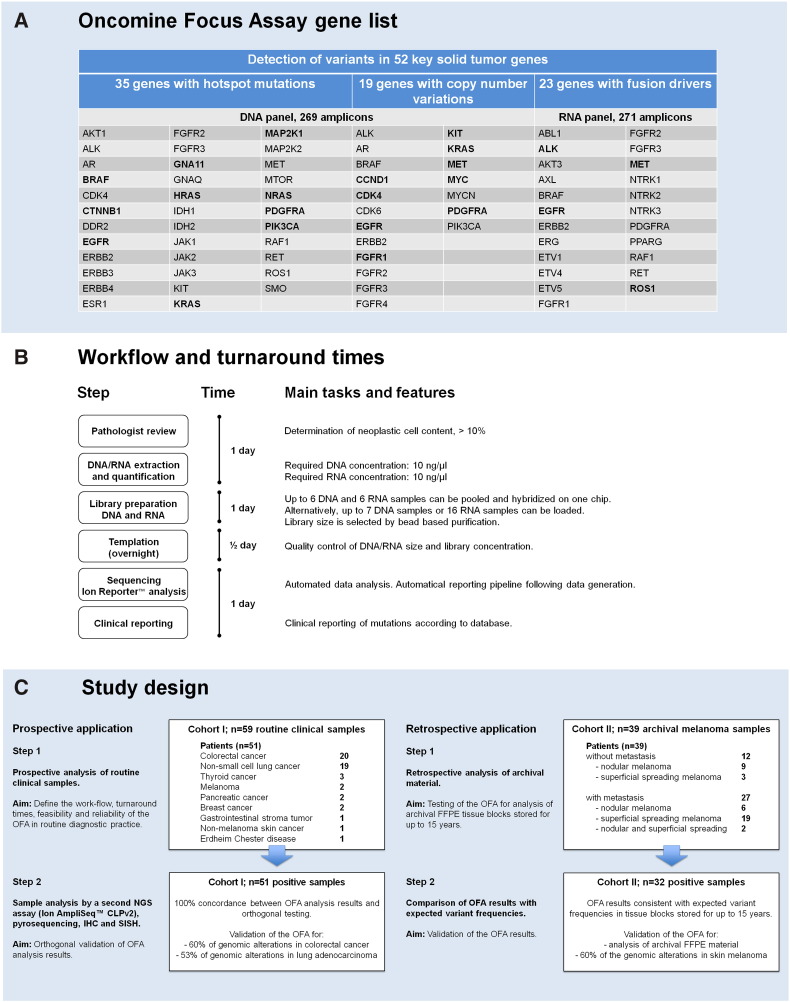
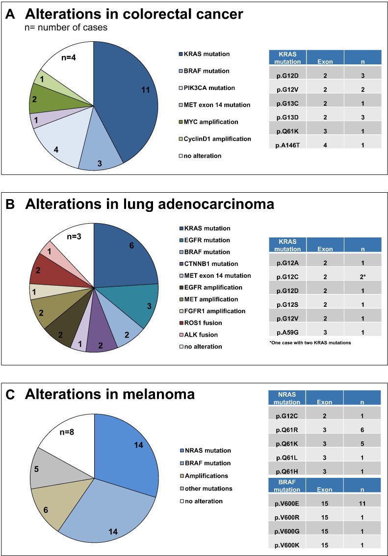
Companion diagnostics rely on genomic testing of molecular alterations to enable effective cancer treatment. Here we report the clinical application and validation of the Oncomine Focus Assay (OFA), an integrated, commercially available next-generation sequencing (NGS) assay for the rapid and simultaneous detection of single nucleotide variants, short insertions and deletions, copy number variations, and gene rearrangements in 52 cancer genes with therapeutic relevance. Two independent patient cohorts were investigated to define the workflow, turnaround times, feasibility, and reliability of OFA targeted sequencing in clinical application and using archival material. Cohort I consisted of 59 diagnostic clinical samples from the daily routine submitted for molecular testing over a 4-month time period. Cohort II consisted of 39 archival melanoma samples that were up to 15years old. Libraries were prepared from isolated nucleic acids and sequenced on the Ion Torrent PGM sequencer. Sequencing datasets were analyzed using the Ion Reporter software. Genomic alterations were identified and validated by orthogonal conventional assays including pyrosequencing and immunohistochemistry. Sequencing results of both cohorts, including archival formalin-fixed, paraffin-embedded material stored up to 15years, were consistent with published variant frequencies. A concordance of 100% between established assays and OFA targeted NGS was observed. The OFA workflow enabled a turnaround of 3½ days. Taken together, OFA was found to be a convenient tool for fast, reliable, broadly applicable and cost-effective targeted NGS of tumor samples in routine diagnostics. Thus, OFA has strong potential to become an important asset for precision oncology.


Figures

Fig. 1
Figure 1
Molecular profiling with the OFA, a targeted multibiomarker NGS assay. (A) The OFA is a targeted NGS panel that includes 52 solid tumor genes associated with current oncology drugs and published evidence. It enables sequencing of 35 hotspot genes, 19 genes associated with copy number gain, and 23 fusion genes, all in a single workflow using the Ion PGM system. Genes printed in bold were detected in cases of the two cohorts studied here. (B) Workflow and turnaround time for molecular profiling of clinical FFPE samples using targeted NGS with the OFA. (C) Design of this study. The OFA was tested and validated on two different cohorts. Cohort I (left) consisted of 59 routine FFPE samples from 51 patients with various solid tumors that were routinely submitted to the molecular pathology service. Half of these samples were randomly selected for orthogonal validation by alternative tests. In addition, validation was performed by comparing the OFA results with expected mutation frequencies from the literature. Cohort II (right) was an archival cohort of 39 FFPE melanoma samples from 39 patients. Mutation frequencies were retrieved from the literature, and the samples of cohort II were analyzed by the OFA retrospectively. The OFA results of the melanomas in cohort II were consistent with the data that can be expected from large published cohorts of cutaneous melanomas.
Fig. 2
Figure 2
Validation of gene fusions and SCNVs detected in routine FFPE samples. (A) EML4-ALK intrachromosomal fusion found in sample no. 32 (NSCLC, adenocarcinoma, H&E left). Confirmation of ALK overexpression due to this EML4-ALK gene fusion by IHC (right). (B) CD74-ROS1 fusion found in sample no. 25 (NSCLC, adenocarcinoma, H&E left). Confirmation of ROS1 overexpression due to this gene fusion between CD74 (Exon 6) and ROS1 (Exon 34) by IHC (right). (C) Intragenic MET fusion (Exon 13-Exon 15; MET “exon 14 skipping”) found in sample no. 29 (lung metastasis of CRC, H&E left). Confirmation of membranous MET overexpression due to this MET exon 14 mutation by IHC (right). (D) MYC amplification found in sample no. 27 (CRC, H&E left). The gene region 8q24.21 on chromosome 8 had a copy number of 25. Confirmation of MYC amplification by IHC (right).
Fig. 3
Figure 3
Genomic aberrations in different tumor types. (A) Actionable variants in 20 CRC patients (n = number of patients) (left). Distribution of KRAS mutations that were detected in 11 (55%) CRC patients (right). (B) Actionable variants detected in 19 NSCLC patients (left). Distribution of KRAS mutations that were detected in six (31.6%) NSCLC patients (right). One NSCLC patient harbored two different KRAS mutations (p. G12C and p. A59G). (C) Actionable variants detected in 39 melanoma patients (left). Distribution of BRAF (right, top panel) and NRAS (right, bottom panel) mutations identified in 14 (35.9%) melanomas.
Fig. 4
Figure 4
Detection and validation of an ALK translocation in one archival melanoma. (A) Relatively symmetrical, exophytic, predominantly intradermal melanocytic tumor with focal epidermal hyperplasia from the right earlobe of a 30-year-old man. (B) Plexiform growth pattern and intersecting fascicles of fusiform melanocytes. Proliferation of spindle and epithelioid melanocytes. (C) The neoplastic melanocytes are positive for ALK in IHC, with strong staining of the cytoplasm. The ALK expression confirms the presence of a TPM3-ALK fusion in this melanoma which was retrospectively reclassified as a melanoma with spitzoid features.

References

    1. Shyr D, Liu Q. Next generation sequencing in cancer research and clinical application. Biol Proced Online. 2013;15:4. - PMC - PubMed
    1. Tripathy D, Harnden K, Blackwell K, Robson M. Next generation sequencing and tumor mutation profiling: are we ready for routine use in the oncology clinic? BMC Med. 2014;12:140. - PMC - PubMed
    1. Strom SP. Current practices and guidelines for clinical next-generation sequencing oncology testing. Cancer Biol Med. 2016;13:3–11. - PMC - PubMed
    1. Luthra R, Chen H, Roy-Chowdhuri S, Singh RR. Next-generation sequencing in clinical molecular diagnostics of cancer: advantages and challenges. Cancers. 2015;7:2023–2036. - PMC - PubMed
    1. Roychowdhury S, Iyer MK, Robinson DR, Lonigro RJ, Wu YM, Cao X, Kalyana-Sundaram S, Sam L, Balbin OA, Quist MJ. Personalized oncology through integrative high-throughput sequencing: a pilot study. Sci Transl Med. 2011;3:111ra121. - PMC - PubMed

LinkOut - more resources