. 2017 Apr 15:53:140-151.
doi: 10.1016/j.actbio.2017.01.068. Epub 2017 Feb 2.

Covalent growth factor tethering to direct neural stem cell differentiation and self-organization

Affiliations

Covalent growth factor tethering to direct neural stem cell differentiation and self-organization

Trevor R Ham et al. Acta Biomater. .

Abstract

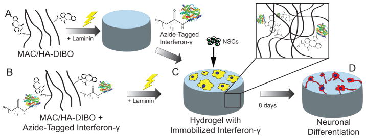
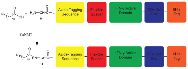
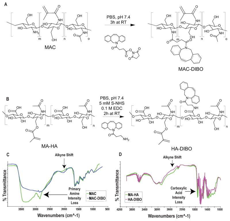
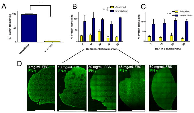
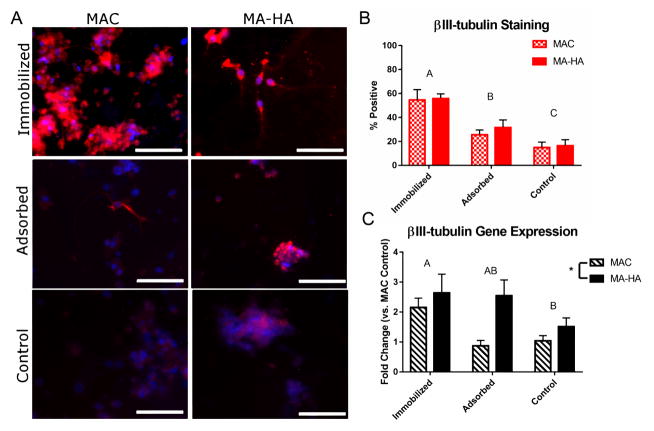
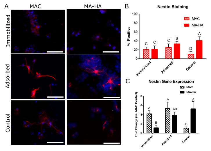
Tethered growth factors offer exciting new possibilities for guiding stem cell behavior. However, many of the current methods present substantial drawbacks which can limit their application and confound results. In this work, we developed a new method for the site-specific covalent immobilization of azide-tagged growth factors and investigated its utility in a model system for guiding neural stem cell (NSC) behavior. An engineered interferon-γ (IFN-γ) fusion protein was tagged with an N-terminal azide group, and immobilized to two different dibenzocyclooctyne-functionalized biomimetic polysaccharides (chitosan and hyaluronan). We successfully immobilized azide-tagged IFN-γ under a wide variety of reaction conditions, both in solution and to bulk hydrogels. To understand the interplay between surface chemistry and protein immobilization, we cultured primary rat NSCs on both materials and showed pronounced biological effects. Expectedly, immobilized IFN-γ increased neuronal differentiation on both materials. Expression of other lineage markers varied depending on the material, suggesting that the interplay of surface chemistry and protein immobilization plays a large role in nuanced cell behavior. We also investigated the bioactivity of immobilized IFN-γ in a 3D environment in vivo and found that it sparked the robust formation of neural tube-like structures from encapsulated NSCs. These findings support a wide range of potential uses for this approach and provide further evidence that adult NSCs are capable of self-organization when exposed to the proper microenvironment.

Statement of significance: For stem cells to be used effectively in regenerative medicine applications, they must be provided with the appropriate cues and microenvironment so that they integrate with existing tissue. This study explores a new method for guiding stem cell behavior: covalent growth factor tethering. We found that adding an N-terminal azide-tag to interferon-γ enabled stable and robust Cu-free 'click' immobilization under a variety of physiologic conditions. We showed that the tagged growth factors retained their bioactivity when immobilized and were able to guide neural stem cell lineage commitment in vitro. We also showed self-organization and neurulation from neural stem cells in vivo. This approach will provide another tool for the orchestration of the complex signaling events required to guide stem cell integration.

Keywords: Central nervous system regeneration; Neural stem cells; Neuroepithelium; Protein immobilization; Strain-promoted alkyne-azide cycloaddition.


Figures

Figure 1
Figure 1
Overview of azIFN-γ ‘click’ immobilization and in vitro NSC differentiation. A) azIFN-γ was immobilized to crosslinked hydrogels (in situ immobilization) or B) immobilized to polymer in solution prior to hydrogel formation (de novo immobilization). C) NSCs were seeded on top of 2D azIFN-γ-functionalized hydrogels where they D) differentiated into neurons after 8 d. Hydrogels were formed from methacrylamide chitosan (MAC) or methacrylated hyaluronan (MA-HA).
Figure 2
Figure 2
azIFN-γ design and production. A recombinant fusion protein was designed, consisting of an azide-tagging sequence, a flexible spacer to allow for proper refolding, the active domain of IFN-γ, a TEV protease cut site (to enable removal of the 6His tag), and a 6His tag for Ni-NTA purification. This protein was N-terminally tagged with 12-ADA to enable SPAAC immobilization.
Figure 3
Figure 3
Synthesis and characterization of MAC-DIBO and HA-DIBO. A) MAC-DIBO was synthesized by reaction of MAC with DIBO-NHS under physiologic conditions. B) HA-DIBO was synthesized by reaction of MA-HA with DIBO-Amine in the presence of S-NHS and EDC as catalysts. C+D) Confirmation of both reactions was verified by FTIR. MAC-DIBO shows a loss of amine intensity and the appearance of an alkyne shift when compared to MAC, while HA-DIBO shows a loss of carboxylic acid intensity and the appearance of an alkyne shift when compared to MA-HA. This indicates the successful functionalization of both MAC and MA-HA with DIBO to enable SPAAC immobilization of azide-tagged IFN-γ.
Figure 4
Figure 4
Immobilization of azIFN-γ through various avenues to chitosan-based (MAC and MAC-DIBO) hydrogels. A) de novo immobilization results in significantly more (≈97%) protein remaining following washing when compared to adsorption (≈3%). The bars show the amount of protein remaining within the gels following washing and enzymatic digestion. B) Differing concentrations of an active protein mixture (FBS) do not affect azIFN-γ immobilization efficiency, while adsorption remains significantly lower. The range from 0 – 60 mg/mL was chosen based on the reported range of physiologic serum protein concentrations,[34] demonstrating the feasibility of our approach within living systems. C) Differing levels of inactive (BSA) protein at supraphysiological levels do not affect azIFN-γ immobilization efficiency, demonstrating the feasibility of our approach within protein-buffered systems. D) Fluorescent images of gels obtained after staining for IFN-γ show an even distribution throughout the gel. The protein was immobilized in situ in the presence of FBS at the concentrations shown in (B). No fluorescence was observed from either primary antibody (no anti-IFN-γ) or protein (no azIFN-γ) negative controls. Scale bars represent 2000 μm. Mean ± SD with n = 4. *** denotes significance (p < 0.001) as determined by two-factor ANOVA with Tukey’s post-hoc.
Figure 5
Figure 5
Neuronal differentiation is caused by immobilized azIFN-γ. A) ICC staining of βIII-tubulin (a key neuronal marker) shows increased expression in immobilized groups for both MAC and MA-HA. Strong neuronal morphologies with longer neurite outgrowth can be seen in the immobilized groups, while the cells in the adsorbed and negative control groups show a more progenitor-like morphology. In general, βIII-tubulin is observed throughout the entire cell body. Red = βIII-tubulin, Blue = Hoechst 33342. Scale bars = 100 m. B) Quantification of ICC results shows a clear trend towards increased βIII-tubulin for immobilized groups over adsorbed and control. The material made no significant difference. Mean ± SD with n = 4. Letters denote significance as determined by two-factor ANOVA with Tukey’s post-hoc. n = 4. C) qPCR analysis shows a similar trend, but with increased expression in the MA-HA adsorbed group. This is likely due to the electrostatic adsorption interaction between MA-HA and IFN-γ, which increased the amount of IFN-γ available to NSCs, but not significantly enough to affect protein expression. Mean ± SE with n = 4, as determined using the method described by Hellemans et al.[32] Letters denote significance as determined by two-factor ANOVA with Tukey’s post-hoc; * denotes significance (p < 0.05) as determined by the same technique.
Figure 6
Figure 6
Immobilized azIFN-γ to MA-HA results in decreased nestin expression, while immobilizing it to MAC results in an increase in expression. A) Staining for nestin reveals cells with neuronal morphologies in the MAC groups while nestin-positive cells in the MA-HA groups appear as immature progenitors. Red = nestin, Blue = Hoechst 33342. Scale bars = 100 m. B) Quantification of ICC imaging shows an unexpected increase in nestin expression when compared with a negative control for MAC groups, while a decrease is seen in MA-HA groups. The increase in nestin expression for MAC groups mirrors the increase in βIII-tubulin staining seen in Figure 5. Mean ± SD with n = 4. Letters denote significance of the interaction term as determined by two-factor ANOVA with Tukey’s post-hoc (p < 0.05). n = 4. C) The same trend is seen in the genetic expression of nestin, with slightly different groupings. These results indicate the possible generation of NeNs from NSCs through the use of MAC. Mean ± SD with n = 4, as determined using the method described by Hellemans et al.[32] Letters denote significance of the interaction term as determined by two-factor ANOVA with Tukey’s post-hoc (p < 0.05).
Figure 7
Figure 7
Robust neural tube-like structures and self-organization from adult NSCs and immobilized azIFN-γ. After maturing 3D MAC-DIBO hydrogel constructs with encapsulated NSCs in rat subcutaneous tissue for 4 weeks, all of the samples containing immobilized azIFN-γ showed self-organized structures throughout sequential sections. The structures are much larger than the pore size of MAC (pores can be easily observed in the βIII-tubulin image). In addition to the expression of neuroepithelium markers, the NSCs can be observed to adopt a polarized orientation in some samples. Scale bars = 100 m.

Similar articles

Cited by

References

    1. Chandra P, Lee SJ. Synthetic extracellular microenvironment for modulating stem cell behaviors. Biomark Insights. 2015;10(Suppl 1):105–16. chandra, Prafulla Lee, Sang Jin ENG Review 2015/06/25 06:00 Biomark Insights 2015 Jun 17 10 Suppl 1 105 16 10.4137/BMI.S20057 eCol- lection 2015. - PMC - PubMed
    1. Illis LS. Central nervous system regeneration does not occur. Spinal Cord. 2012;50(4):259–63. doi: 10.1038/sc.2011.132. - DOI - PubMed
    1. Li H, Koenig AM, Sloan P, Leipzig ND. In vivo assessment of guided neural stem cell differentiation in growth factor immobilized chitosan-based hydrogel scaffolds. Biomaterials. 2014;35(33):9049–57. doi: 10.1016/j.biomaterials.2014.07.038. - DOI - PubMed
    1. Aertker BM, Bedi S, Cox JCS. Strategies for cns repair following tbi. Exp Neurol. 2016;275(Pt 3):411–26. doi: 10.1016/j.expneurol.2015.01.008. - DOI - PubMed
    1. Sarhan F, Saif D, Saif A. An overview of traumatic spinal cord injury: part 1. aetiology and pathophysiology. British Journal of Neuroscience Nursing. 2012;8(6):319–325.

Publication types

MeSH terms

LinkOut - more resources